

contribute. In the following, I will leave aside the biological implications of the works presented,
which can be fascinating but are beyond the scope of the present text, and try to extract from published
papers technical information that can be relevant to the use of APols for EM studies of other biological
systems. Table 12.1 lists the 27 MP studies that had come to my attention by the time the writing of this
chapter was completed, plus one study (Study 12.20) on a non-membrane system that may shed some
light on what is going on with MPs. The speed at which SP-EM studies of APol-trapped MPs are
published has become very rapid. A dozen more studies that were published or came to my attention
too late to be included in the table and discussions are mentioned at the beginning of § 12.3.2
(NS studies) and the end of 12.3.3 (cryo-EM studies). In the following, biological background and
experimental details are kept to a minimum except when they appear particularly relevant to a rational
use of APols. A quick summary of each study and some complementary information, such as the
molecular masses of the complexes studied, can be found in the table.
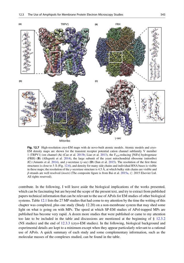
Fig. 12.7 High-resolution cryo-EM maps with de novo-built atomic models. Atomic models and cryoEM density maps are shown for the transient receptor potential cation channel subfamily V member
1 (TRPV1) ion channel (A) (Cao et al. 2013b; Liao et al. 2013), the F 420 -reducing [NiFe] hydrogenase
(FRH) (B) (Allegretti et al. 2014), the large subunit of the yeast mitochondrial ribosome (mitoribo)
(C) (Amunts et al. 2014), and γ-secretase (γ-sec) (D) (Sun et al. 2015). The resolution of the first three
structures is close to 3 Å (Fig. 12.6), and density for many side chains and individual RNA bases is visible
in these maps; the resolution of the γ-secretase structure is 4.5 Å, at which bulky side chains are visible and
β-strands are well resolved (insets) (The composite figure is from Bai et al. 2015a, # 2015 Elsevier Ltd.
All rights reserved).
12.3 The Use of Amphipols for Membrane Protein Electron Microscopy Studies
543
which can be fascinating but are beyond the scope of the present text, and try to extract from published
papers technical information that can be relevant to the use of APols for EM studies of other biological
systems. Table 12.1 lists the 27 MP studies that had come to my attention by the time the writing of this
chapter was completed, plus one study (Study 12.20) on a non-membrane system that may shed some
light on what is going on with MPs. The speed at which SP-EM studies of APol-trapped MPs are
published has become very rapid. A dozen more studies that were published or came to my attention
too late to be included in the table and discussions are mentioned at the beginning of § 12.3.2
(NS studies) and the end of 12.3.3 (cryo-EM studies). In the following, biological background and
experimental details are kept to a minimum except when they appear particularly relevant to a rational
use of APols. A quick summary of each study and some complementary information, such as the
molecular masses of the complexes studied, can be found in the table.
Fig. 12.7 High-resolution cryo-EM maps with de novo-built atomic models. Atomic models and cryoEM density maps are shown for the transient receptor potential cation channel subfamily V member
1 (TRPV1) ion channel (A) (Cao et al. 2013b; Liao et al. 2013), the F 420 -reducing [NiFe] hydrogenase
(FRH) (B) (Allegretti et al. 2014), the large subunit of the yeast mitochondrial ribosome (mitoribo)
(C) (Amunts et al. 2014), and γ-secretase (γ-sec) (D) (Sun et al. 2015). The resolution of the first three
structures is close to 3 Å (Fig. 12.6), and density for many side chains and individual RNA bases is visible
in these maps; the resolution of the γ-secretase structure is 4.5 Å, at which bulky side chains are visible and
β-strands are well resolved (insets) (The composite figure is from Bai et al. 2015a, # 2015 Elsevier Ltd.
All rights reserved).
12.3 The Use of Amphipols for Membrane Protein Electron Microscopy Studies
543