

studies of the factors that come into play and can make APols useful or useless auxiliaries for EM
studies. Second, APols are often considered by electron microscopists as just an alternative “detergent,” and no specific mention is made of their use in abstracts or keywords, nor, in many cases, is the
APol literature referred to. A bibliographical search based on titles, keywords, or literature cited can
therefore miss interesting studies – hence the absence of references to the Wilkens/Capaldi work on the
F 1 F O ATP synthase (Study 12.2 in Table 12.1) in our early general reviews on APols and their
applications. In the following, I have done my best to offer an exhaustive survey of relevant
publications, but I cannot guarantee that some potentially instructive studies have not been overlooked.
EM studies of MPs stabilized by APols have yielded much more novel biological information
than have, to date, NMR or crystallographic studies. As a result, authors have tended to focus on the
biological implications of their work and to spend little time on experimental details, or on the
discussion of what APols contributed to their work and why. The description of experimental methods
is often skimpy, with the source and exact nature of the APol used sometimes remaining unspecified
and the protocol of transfer not described in detail. One can be left uncertain, for instance, whether free
APol was separated from the complexes before EM grids were prepared, or about how much time
elapsed between blotting the EM grid and freezing it, even though these can be critical parameters
influencing the thickness of the film and particle dispersity (see below, § 12.3.4.2). This dearth of data
is much to be regretted, because it complicates the interpretation of the results and slows down the
identification of pitfalls and good practices in a field to which APols appear to have so much to
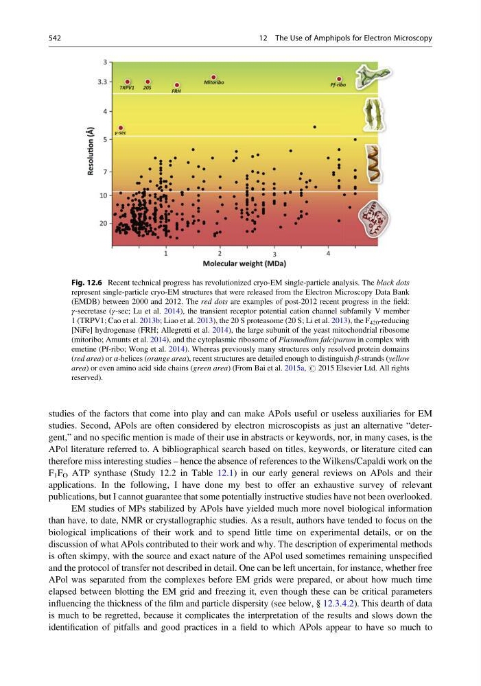
Fig. 12.6 Recent technical progress has revolutionized cryo-EM single-particle analysis. The black dots
represent single-particle cryo-EM structures that were released from the Electron Microscopy Data Bank
(EMDB) between 2000 and 2012. The red dots are examples of post-2012 recent progress in the field:
γ-secretase (γ-sec; Lu et al. 2014), the transient receptor potential cation channel subfamily V member
1 (TRPV1; Cao et al. 2013b; Liao et al. 2013), the 20 S proteasome (20 S; Li et al. 2013), the F 420 -reducing
[NiFe] hydrogenase (FRH; Allegretti et al. 2014), the large subunit of the yeast mitochondrial ribosome
(mitoribo; Amunts et al. 2014), and the cytoplasmic ribosome of Plasmodium falciparum in complex with
emetine (Pf-ribo; Wong et al. 2014). Whereas previously many structures only resolved protein domains
(red area) or α-helices (orange area), recent structures are detailed enough to distinguish β-strands (yellow
area) or even amino acid side chains (green area) (From Bai et al. 2015a, # 2015 Elsevier Ltd. All rights
reserved).
542
12 The Use of Amphipols for Electron Microscopy
studies. Second, APols are often considered by electron microscopists as just an alternative “detergent,” and no specific mention is made of their use in abstracts or keywords, nor, in many cases, is the
APol literature referred to. A bibliographical search based on titles, keywords, or literature cited can
therefore miss interesting studies – hence the absence of references to the Wilkens/Capaldi work on the
F 1 F O ATP synthase (Study 12.2 in Table 12.1) in our early general reviews on APols and their
applications. In the following, I have done my best to offer an exhaustive survey of relevant
publications, but I cannot guarantee that some potentially instructive studies have not been overlooked.
EM studies of MPs stabilized by APols have yielded much more novel biological information
than have, to date, NMR or crystallographic studies. As a result, authors have tended to focus on the
biological implications of their work and to spend little time on experimental details, or on the
discussion of what APols contributed to their work and why. The description of experimental methods
is often skimpy, with the source and exact nature of the APol used sometimes remaining unspecified
and the protocol of transfer not described in detail. One can be left uncertain, for instance, whether free
APol was separated from the complexes before EM grids were prepared, or about how much time
elapsed between blotting the EM grid and freezing it, even though these can be critical parameters
influencing the thickness of the film and particle dispersity (see below, § 12.3.4.2). This dearth of data
is much to be regretted, because it complicates the interpretation of the results and slows down the
identification of pitfalls and good practices in a field to which APols appear to have so much to
Fig. 12.6 Recent technical progress has revolutionized cryo-EM single-particle analysis. The black dots
represent single-particle cryo-EM structures that were released from the Electron Microscopy Data Bank
(EMDB) between 2000 and 2012. The red dots are examples of post-2012 recent progress in the field:
γ-secretase (γ-sec; Lu et al. 2014), the transient receptor potential cation channel subfamily V member
1 (TRPV1; Cao et al. 2013b; Liao et al. 2013), the 20 S proteasome (20 S; Li et al. 2013), the F 420 -reducing
[NiFe] hydrogenase (FRH; Allegretti et al. 2014), the large subunit of the yeast mitochondrial ribosome
(mitoribo; Amunts et al. 2014), and the cytoplasmic ribosome of Plasmodium falciparum in complex with
emetine (Pf-ribo; Wong et al. 2014). Whereas previously many structures only resolved protein domains
(red area) or α-helices (orange area), recent structures are detailed enough to distinguish β-strands (yellow
area) or even amino acid side chains (green area) (From Bai et al. 2015a, # 2015 Elsevier Ltd. All rights
reserved).
542
12 The Use of Amphipols for Electron Microscopy