

(iii) Uneven dispersion of the particles, which tend to be pushed away from the thin central
region of the aqueous film and to crowd in the meniscus at the rim of the holes, where they
lie in a thicker region of the film, lowering the contrast, and tend to interact with one another
(see e.g. Flötenmeyer et al. 2007; Baker et al. 2015; Jeong et al. 2016; cf. below Figs. 12.9
and 12.26).
It could be hoped that APols would help with the first two problems, because (i) they tend to
stabilize most MPs and MP complexes as compared to detergent solutions, and (ii) free APols do not need
to be present in high concentration, and can even be totally removed if the risk of a degree of aggregation
is assumed (see Chap. 5). It turned out that, unexpectedly, APols also seem to help with problem (iii).
12.3 The Use of Amphipols for Membrane Protein Electron Microscopy
Studies
The literature in the field is somewhat hard to follow and analyze, for two reasons. First, at variance
with other structural approaches such as NMR and crystallography, there have been few systematic
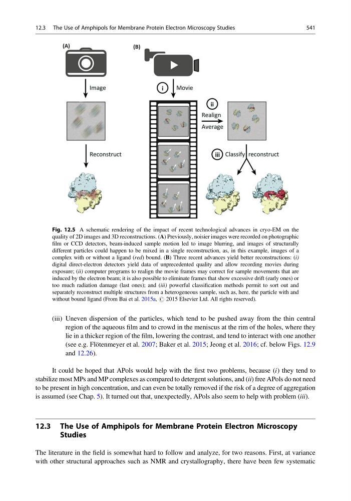
Fig. 12.5 A schematic rendering of the impact of recent technological advances in cryo-EM on the
quality of 2D images and 3D reconstructions. (A) Previously, noisier images were recorded on photographic
film or CCD detectors, beam-induced sample motion led to image blurring, and images of structurally
different particles could happen to be mixed in a single reconstruction, as, in this example, images of a
complex with or without a ligand (red) bound. (B) Three recent advances yield better reconstructions: (i)
digital direct-electron detectors yield data of unprecedented quality and allow recording movies during
exposure; (ii) computer programs to realign the movie frames may correct for sample movements that are
induced by the electron beam; it is also possible to eliminate frames that show excessive drift (early ones) or
too much radiation damage (last ones); and (iii) powerful classification methods permit to sort out and
separately reconstruct multiple structures from a heterogeneous sample, such as, here, the particle with and
without bound ligand (From Bai et al. 2015a, # 2015 Elsevier Ltd. All rights reserved).
12.3 The Use of Amphipols for Membrane Protein Electron Microscopy Studies
541
region of the aqueous film and to crowd in the meniscus at the rim of the holes, where they
lie in a thicker region of the film, lowering the contrast, and tend to interact with one another
(see e.g. Flötenmeyer et al. 2007; Baker et al. 2015; Jeong et al. 2016; cf. below Figs. 12.9
and 12.26).
It could be hoped that APols would help with the first two problems, because (i) they tend to
stabilize most MPs and MP complexes as compared to detergent solutions, and (ii) free APols do not need
to be present in high concentration, and can even be totally removed if the risk of a degree of aggregation
is assumed (see Chap. 5). It turned out that, unexpectedly, APols also seem to help with problem (iii).
12.3 The Use of Amphipols for Membrane Protein Electron Microscopy
Studies
The literature in the field is somewhat hard to follow and analyze, for two reasons. First, at variance
with other structural approaches such as NMR and crystallography, there have been few systematic
Fig. 12.5 A schematic rendering of the impact of recent technological advances in cryo-EM on the
quality of 2D images and 3D reconstructions. (A) Previously, noisier images were recorded on photographic
film or CCD detectors, beam-induced sample motion led to image blurring, and images of structurally
different particles could happen to be mixed in a single reconstruction, as, in this example, images of a
complex with or without a ligand (red) bound. (B) Three recent advances yield better reconstructions: (i)
digital direct-electron detectors yield data of unprecedented quality and allow recording movies during
exposure; (ii) computer programs to realign the movie frames may correct for sample movements that are
induced by the electron beam; it is also possible to eliminate frames that show excessive drift (early ones) or
too much radiation damage (last ones); and (iii) powerful classification methods permit to sort out and
separately reconstruct multiple structures from a heterogeneous sample, such as, here, the particle with and
without bound ligand (From Bai et al. 2015a, # 2015 Elsevier Ltd. All rights reserved).
12.3 The Use of Amphipols for Membrane Protein Electron Microscopy Studies
541