

12.2.4 Single-Particle Electron Microscopy Studies of Membrane Proteins
Cryo-EM studies of MPs present special challenges, essentially due to the presence of detergent.
Among difficulties attributed to the detergent are the following:
(i) Biochemical instability of the target protein (see Chap. 2).
(ii) Loss of contrast (cf. Mazhab-Jafari and Rubinstein 2016; Stark and Chari 2016); this is
probably not a serious problem with detergents with a low critical micellar concentration
(CMC), like maltose neopentyl glycol (MNG) detergents or digitonin, which do not need to
be present at a high concentration for MPs to remain soluble, but it can become one with
detergents like CHAPS or CHAPSO, whose concentration must be kept above their high
CMC of ~8 mM (~5 gÁL
À1 ).
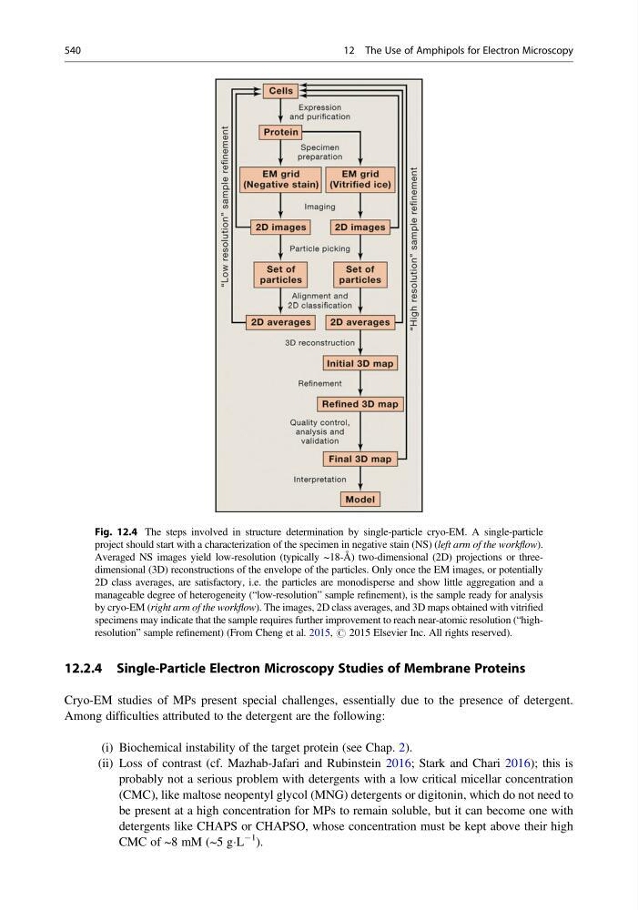
Fig. 12.4 The steps involved in structure determination by single-particle cryo-EM. A single-particle
project should start with a characterization of the specimen in negative stain (NS) (left arm of the workflow).
Averaged NS images yield low-resolution (typically ~18-Å) two-dimensional (2D) projections or threedimensional (3D) reconstructions of the envelope of the particles. Only once the EM images, or potentially
2D class averages, are satisfactory, i.e. the particles are monodisperse and show little aggregation and a
manageable degree of heterogeneity (“low-resolution” sample refinement), is the sample ready for analysis
by cryo-EM (right arm of the workflow). The images, 2D class averages, and 3D maps obtained with vitrified
specimens may indicate that the sample requires further improvement to reach near-atomic resolution (“highresolution” sample refinement) (From Cheng et al. 2015, # 2015 Elsevier Inc. All rights reserved).
540
12 The Use of Amphipols for Electron Microscopy
Cryo-EM studies of MPs present special challenges, essentially due to the presence of detergent.
Among difficulties attributed to the detergent are the following:
(i) Biochemical instability of the target protein (see Chap. 2).
(ii) Loss of contrast (cf. Mazhab-Jafari and Rubinstein 2016; Stark and Chari 2016); this is
probably not a serious problem with detergents with a low critical micellar concentration
(CMC), like maltose neopentyl glycol (MNG) detergents or digitonin, which do not need to
be present at a high concentration for MPs to remain soluble, but it can become one with
detergents like CHAPS or CHAPSO, whose concentration must be kept above their high
CMC of ~8 mM (~5 gÁL
À1 ).
Fig. 12.4 The steps involved in structure determination by single-particle cryo-EM. A single-particle
project should start with a characterization of the specimen in negative stain (NS) (left arm of the workflow).
Averaged NS images yield low-resolution (typically ~18-Å) two-dimensional (2D) projections or threedimensional (3D) reconstructions of the envelope of the particles. Only once the EM images, or potentially
2D class averages, are satisfactory, i.e. the particles are monodisperse and show little aggregation and a
manageable degree of heterogeneity (“low-resolution” sample refinement), is the sample ready for analysis
by cryo-EM (right arm of the workflow). The images, 2D class averages, and 3D maps obtained with vitrified
specimens may indicate that the sample requires further improvement to reach near-atomic resolution (“highresolution” sample refinement) (From Cheng et al. 2015, # 2015 Elsevier Inc. All rights reserved).
540
12 The Use of Amphipols for Electron Microscopy