

for hours at a time in order to collect the massive amounts of data required. This progress, which makes
it possible to collect and selectively average those images that are least affected by radiation damage
and drift, has had the dramatic consequence that 3D structures derived from single-particle cryo-EM
images have now reached or exceeded a resolution of ~4 Å. At this critical resolution, bulky amino
acid side chains can be identified, and it becomes possible to directly fit atomic models into density
maps, without having to rely on extraneous data such as X-ray models of individual subunits or
domains (see Figs. 12.1, 12.6, and 12.7).
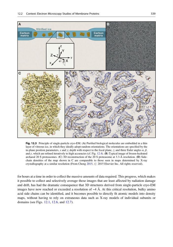
Fig. 12.3 Principle of single-particle cryo-EM. (A) Purified biological molecules are embedded in a thin
layer of vitreous ice, in which they ideally adopt random orientations. The orientations are specified by the
in-plane position parameters, x and y; depth with respect to the focal plane, z; and three Euler angles α, β,
and γ, which are refined iteratively to high accuracies (cf. Fig. 12.4). (B) Typical image of frozen-hydrated
archaeal 20 S proteasomes. (C) 3D reconstruction of the 20 S proteasome at 3.3-Å resolution. (D) Sidechain densities of the map shown in C are comparable to those seen in maps determined by X-ray
crystallography at a similar resolution (From Cheng 2015, # 2015 Elsevier Inc. All rights reserved).
12.2 Context: Electron Microscopy Studies of Membrane Proteins
539
it possible to collect and selectively average those images that are least affected by radiation damage
and drift, has had the dramatic consequence that 3D structures derived from single-particle cryo-EM
images have now reached or exceeded a resolution of ~4 Å. At this critical resolution, bulky amino
acid side chains can be identified, and it becomes possible to directly fit atomic models into density
maps, without having to rely on extraneous data such as X-ray models of individual subunits or
domains (see Figs. 12.1, 12.6, and 12.7).
Fig. 12.3 Principle of single-particle cryo-EM. (A) Purified biological molecules are embedded in a thin
layer of vitreous ice, in which they ideally adopt random orientations. The orientations are specified by the
in-plane position parameters, x and y; depth with respect to the focal plane, z; and three Euler angles α, β,
and γ, which are refined iteratively to high accuracies (cf. Fig. 12.4). (B) Typical image of frozen-hydrated
archaeal 20 S proteasomes. (C) 3D reconstruction of the 20 S proteasome at 3.3-Å resolution. (D) Sidechain densities of the map shown in C are comparable to those seen in maps determined by X-ray
crystallography at a similar resolution (From Cheng 2015, # 2015 Elsevier Inc. All rights reserved).
12.2 Context: Electron Microscopy Studies of Membrane Proteins
539