

Box 12.1 (continued)
12.2.3 Single-Particle Cryo-EM Studies of Biological Molecules:
The Resolution Revolution
It is no exaggeration to say that, since 2012, the field of single-particle cryo-EM has undergone a
revolution (for analyses and comments, see e.g. Henderson 2013, 2015; Agard et al. 2014; Kühlbrandt
2014a, b; Liao et al. 2014; Bai et al. 2015a; Callaway 2015; Cheng 2015; Schröder 2015; Vinothkumar
2015; De Zorzi et al. 2016; Vinothkumar and Henderson 2016). This is principally due to two main
factors, which combine to produce much better images. First, direct-electron detector device (DDD)
cameras have been developed, which, not having to convert electrons into photons to be able to detect
them, have a higher detective quantum efficiency and better signal-to-noise ratio than the CCD
cameras used previously. Second, the underlying complementary metal-oxide semiconductor
(CMOS) technology makes it possible to collect series of views of a given field during illumination,
rather than a single averaged one (Fig. 12.5Bi). This permits to implement much more efficient
corrections of the movements that the specimen undergoes under the electron beam (due, in particular,
to annealing of the stresses in the vitrified water film) (Fig. 12.5Bii). The higher quality of the images,
in turn (cf. Fig. 12.1C), improves the performances of the alignment and 3D reconstruction programs,
which, themselves, have undergone considerable progress made implementable by the constant
increase in computing power. Also of significant help are advances in computer control of EM
multi-specimen handling and stage movement, which allow “driverless” running of the microscope
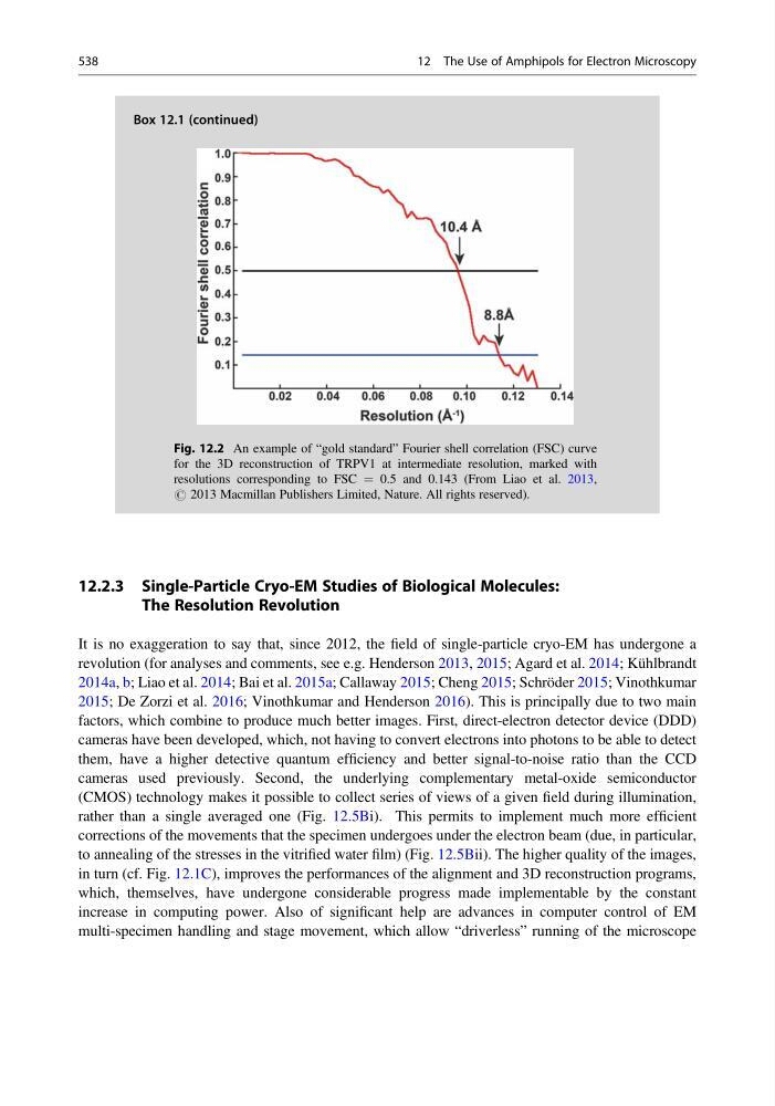
Fig. 12.2 An example of “gold standard” Fourier shell correlation (FSC) curve
for the 3D reconstruction of TRPV1 at intermediate resolution, marked with
resolutions corresponding to FSC ¼ 0.5 and 0.143 (From Liao et al. 2013,
# 2013 Macmillan Publishers Limited, Nature. All rights reserved).
538
12 The Use of Amphipols for Electron Microscopy
12.2.3 Single-Particle Cryo-EM Studies of Biological Molecules:
The Resolution Revolution
It is no exaggeration to say that, since 2012, the field of single-particle cryo-EM has undergone a
revolution (for analyses and comments, see e.g. Henderson 2013, 2015; Agard et al. 2014; Kühlbrandt
2014a, b; Liao et al. 2014; Bai et al. 2015a; Callaway 2015; Cheng 2015; Schröder 2015; Vinothkumar
2015; De Zorzi et al. 2016; Vinothkumar and Henderson 2016). This is principally due to two main
factors, which combine to produce much better images. First, direct-electron detector device (DDD)
cameras have been developed, which, not having to convert electrons into photons to be able to detect
them, have a higher detective quantum efficiency and better signal-to-noise ratio than the CCD
cameras used previously. Second, the underlying complementary metal-oxide semiconductor
(CMOS) technology makes it possible to collect series of views of a given field during illumination,
rather than a single averaged one (Fig. 12.5Bi). This permits to implement much more efficient
corrections of the movements that the specimen undergoes under the electron beam (due, in particular,
to annealing of the stresses in the vitrified water film) (Fig. 12.5Bii). The higher quality of the images,
in turn (cf. Fig. 12.1C), improves the performances of the alignment and 3D reconstruction programs,
which, themselves, have undergone considerable progress made implementable by the constant
increase in computing power. Also of significant help are advances in computer control of EM
multi-specimen handling and stage movement, which allow “driverless” running of the microscope
Fig. 12.2 An example of “gold standard” Fourier shell correlation (FSC) curve
for the 3D reconstruction of TRPV1 at intermediate resolution, marked with
resolutions corresponding to FSC ¼ 0.5 and 0.143 (From Liao et al. 2013,
# 2013 Macmillan Publishers Limited, Nature. All rights reserved).
538
12 The Use of Amphipols for Electron Microscopy