

Box 12.1 Fourier Transforms, Thon Rings, and Fourier Shell Correlation
The Fourier transform is a mathematical operation that decomposes a function of time or space
(a signal) into the frequencies that make it up. Fourier transforms are used widely in biophysics, in
domains as varied as crystallography, small-angle radiation scattering, NMR, and infrared spectroscopy. Very often, the signal that is recorded by the experimenter is the Fourier transform of
some property of the object under study, such as the distribution of electron density. In X-ray
or neutron crystallography, the diffraction patterns are the transform of the electron or neutron
scattering length density distribution in the crystals, in electron crystallography that of the potential
map. They are formed of discrete peaks or rings because of the crystalline nature of the sample.
EM images of 2D crystals are Fourier-transformed mathematically in order to exploit the crystalline
nature of the sample to extract signals from the noise. In small-angle X-ray or neutron scattering, the
sample is disordered and the signal measured is a continuous Fourier transform, which, at small
angles, contains information about long-distance correlations, such as the distribution of molecules
in the solution (attraction, repulsion, random distribution. . .), at larger angles, about their size and
mass, and, at still larger angles, about their shape and internal structure (see Chap. 9, Box 9.2).
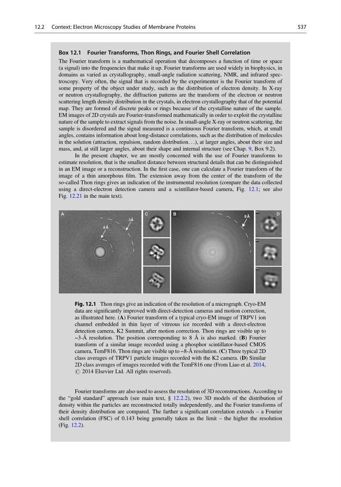
In the present chapter, we are mostly concerned with the use of Fourier transforms to
estimate resolution, that is the smallest distance between structural details that can be distinguished
in an EM image or a reconstruction. In the first case, one can calculate a Fourier transform of the
image of a thin amorphous film. The extension away from the center of the transform of the
so-called Thon rings gives an indication of the instrumental resolution (compare the data collected
using a direct-electron detection camera and a scintillator-based camera, Fig. 12.1; see also
Fig. 12.21 in the main text).
Fourier transforms are also used to assess the resolution of 3D reconstructions. According to
the “gold standard” approach (see main text, § 12.2.2), two 3D models of the distribution of
density within the particles are reconstructed totally independently, and the Fourier transforms of
their density distribution are compared. The farther a significant correlation extends – a Fourier
shell correlation (FSC) of 0.143 being generally taken as the limit – the higher the resolution
(Fig. 12.2).
Fig. 12.1 Thon rings give an indication of the resolution of a micrograph. Cryo-EM
data are significantly improved with direct-detection cameras and motion correction,
as illustrated here. (A) Fourier transform of a typical cryo-EM image of TRPV1 ion
channel embedded in thin layer of vitreous ice recorded with a direct-electron
detection camera, K2 Summit, after motion correction. Thon rings are visible up to
~3-Å resolution. The position corresponding to 8 Å is also marked. (B) Fourier
transform of a similar image recorded using a phosphor scintillator-based CMOS
camera, TemF816. Thon rings are visible up to ~8-Å resolution. (C) Three typical 2D
class averages of TRPV1 particle images recorded with the K2 camera. (D) Similar
2D class averages of images recorded with the TemF816 one (From Liao et al. 2014,
# 2014 Elsevier Ltd. All rights reserved).
12.2 Context: Electron Microscopy Studies of Membrane Proteins
537
The Fourier transform is a mathematical operation that decomposes a function of time or space
(a signal) into the frequencies that make it up. Fourier transforms are used widely in biophysics, in
domains as varied as crystallography, small-angle radiation scattering, NMR, and infrared spectroscopy. Very often, the signal that is recorded by the experimenter is the Fourier transform of
some property of the object under study, such as the distribution of electron density. In X-ray
or neutron crystallography, the diffraction patterns are the transform of the electron or neutron
scattering length density distribution in the crystals, in electron crystallography that of the potential
map. They are formed of discrete peaks or rings because of the crystalline nature of the sample.
EM images of 2D crystals are Fourier-transformed mathematically in order to exploit the crystalline
nature of the sample to extract signals from the noise. In small-angle X-ray or neutron scattering, the
sample is disordered and the signal measured is a continuous Fourier transform, which, at small
angles, contains information about long-distance correlations, such as the distribution of molecules
in the solution (attraction, repulsion, random distribution. . .), at larger angles, about their size and
mass, and, at still larger angles, about their shape and internal structure (see Chap. 9, Box 9.2).
In the present chapter, we are mostly concerned with the use of Fourier transforms to
estimate resolution, that is the smallest distance between structural details that can be distinguished
in an EM image or a reconstruction. In the first case, one can calculate a Fourier transform of the
image of a thin amorphous film. The extension away from the center of the transform of the
so-called Thon rings gives an indication of the instrumental resolution (compare the data collected
using a direct-electron detection camera and a scintillator-based camera, Fig. 12.1; see also
Fig. 12.21 in the main text).
Fourier transforms are also used to assess the resolution of 3D reconstructions. According to
the “gold standard” approach (see main text, § 12.2.2), two 3D models of the distribution of
density within the particles are reconstructed totally independently, and the Fourier transforms of
their density distribution are compared. The farther a significant correlation extends – a Fourier
shell correlation (FSC) of 0.143 being generally taken as the limit – the higher the resolution
(Fig. 12.2).
Fig. 12.1 Thon rings give an indication of the resolution of a micrograph. Cryo-EM
data are significantly improved with direct-detection cameras and motion correction,
as illustrated here. (A) Fourier transform of a typical cryo-EM image of TRPV1 ion
channel embedded in thin layer of vitreous ice recorded with a direct-electron
detection camera, K2 Summit, after motion correction. Thon rings are visible up to
~3-Å resolution. The position corresponding to 8 Å is also marked. (B) Fourier
transform of a similar image recorded using a phosphor scintillator-based CMOS
camera, TemF816. Thon rings are visible up to ~8-Å resolution. (C) Three typical 2D
class averages of TRPV1 particle images recorded with the K2 camera. (D) Similar
2D class averages of images recorded with the TemF816 one (From Liao et al. 2014,
# 2014 Elsevier Ltd. All rights reserved).
12.2 Context: Electron Microscopy Studies of Membrane Proteins
537