

ratio of ~5%. Increasing the APol/MO mass ratio above 7% induces a transition to a cubic phase with
Ia3d symmetry, and then the phase transforms to a lamellar phase at mass ratios higher than 20%
(Sintsov et al. unpublished results). This is likely the reason why no high-quality BR crystals were
observed in the presence of an excess of APol. Indeed, under the conditions used, the APol/MO mass
ratio was ~12%, which is above the ratio compatible with a Pn3m cubic phase.
As already mentioned, removal of free APol from preparations of MP/APol complexes, including BR/A8-35 ones, leads to a variable degree of aggregation (Zoonens et al. 2007; Gohon et al. 2008;
Arunmanee et al. 2014; Charvolin et al. 2014) (cf. Chaps. 5, § 5.2.1 and 12, § 12.3). Indeed, after
removal of free APol, the main peak observed in the size exclusion chromatography elution profile of
the BR/A8-35 complexes became slightly broader and eluted slightly earlier than before removal
(Polovinkin et al. 2014). This behavior is similar to that observed with tOmpA/A8-35 complexes
(Zoonens et al. 2007) (see Chap. 5, Fig. 5.6). It reflects the formation of small oligomers, as well as a
very small amount of larger aggregates, which elute in the void volume. At the time of mixing with
MO, BR/A8-35 complexes depleted of free APol therefore are not homogeneous. This does not
prevent BR from reorganizing into well-ordered 3D crystals once inserted into the lipidic mesophase.
Similarly, in the presence of lipopolysaccharide, OmpF/A8-35 filaments can evolve toward 2D crystals
(Arunmanee et al. 2014).
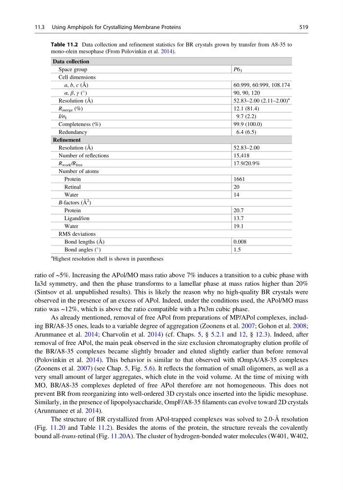
The structure of BR crystallized from APol-trapped complexes was solved to 2.0-Å resolution
(Fig. 11.20 and Table 11.2). Besides the atoms of the protein, the structure reveals the covalently
bound all-trans-retinal (Fig. 11.20A). The cluster of hydrogen-bonded water molecules (W401, W402,
Table 11.2 Data collection and refinement statistics for BR crystals grown by transfer from A8-35 to
mono-olein mesophase (From Polovinkin et al. 2014).
Data collection
Space group
P6 3
Cell dimensions
a, b, c (Å)
60.999, 60.999, 108.174
α, β, γ (
)
90, 90, 120
Resolution (Å)
52.83–2.00 (2.11–2.00)
a
R merge (%)
12.1 (81.4)
I/σ I
9.7 (2.2)
Completeness (%)
99.9 (100.0)
Redundancy
6.4 (6.5)
Refinement
Resolution (Å)
52.83–2.00
Number of reflections
15,418
R work /R free
17.9/20.9%
Number of atoms
Protein
1661
Retinal
20
Water
14
B-factors (Å
2
)
Protein
20.7
Ligand/ion
13.7
Water
19.1
RMS deviations
Bond lengths (Å)
0.008
Bond angles (
)
1.5
a Highest resolution shell is shown in parentheses
11.3 Using Amphipols for Crystallizing Membrane Proteins
519
Ia3d symmetry, and then the phase transforms to a lamellar phase at mass ratios higher than 20%
(Sintsov et al. unpublished results). This is likely the reason why no high-quality BR crystals were
observed in the presence of an excess of APol. Indeed, under the conditions used, the APol/MO mass
ratio was ~12%, which is above the ratio compatible with a Pn3m cubic phase.
As already mentioned, removal of free APol from preparations of MP/APol complexes, including BR/A8-35 ones, leads to a variable degree of aggregation (Zoonens et al. 2007; Gohon et al. 2008;
Arunmanee et al. 2014; Charvolin et al. 2014) (cf. Chaps. 5, § 5.2.1 and 12, § 12.3). Indeed, after
removal of free APol, the main peak observed in the size exclusion chromatography elution profile of
the BR/A8-35 complexes became slightly broader and eluted slightly earlier than before removal
(Polovinkin et al. 2014). This behavior is similar to that observed with tOmpA/A8-35 complexes
(Zoonens et al. 2007) (see Chap. 5, Fig. 5.6). It reflects the formation of small oligomers, as well as a
very small amount of larger aggregates, which elute in the void volume. At the time of mixing with
MO, BR/A8-35 complexes depleted of free APol therefore are not homogeneous. This does not
prevent BR from reorganizing into well-ordered 3D crystals once inserted into the lipidic mesophase.
Similarly, in the presence of lipopolysaccharide, OmpF/A8-35 filaments can evolve toward 2D crystals
(Arunmanee et al. 2014).
The structure of BR crystallized from APol-trapped complexes was solved to 2.0-Å resolution
(Fig. 11.20 and Table 11.2). Besides the atoms of the protein, the structure reveals the covalently
bound all-trans-retinal (Fig. 11.20A). The cluster of hydrogen-bonded water molecules (W401, W402,
Table 11.2 Data collection and refinement statistics for BR crystals grown by transfer from A8-35 to
mono-olein mesophase (From Polovinkin et al. 2014).
Data collection
Space group
P6 3
Cell dimensions
a, b, c (Å)
60.999, 60.999, 108.174
α, β, γ (
)
90, 90, 120
Resolution (Å)
52.83–2.00 (2.11–2.00)
a
R merge (%)
12.1 (81.4)
I/σ I
9.7 (2.2)
Completeness (%)
99.9 (100.0)
Redundancy
6.4 (6.5)
Refinement
Resolution (Å)
52.83–2.00
Number of reflections
15,418
R work /R free
17.9/20.9%
Number of atoms
Protein
1661
Retinal
20
Water
14
B-factors (Å
2
)
Protein
20.7
Ligand/ion
13.7
Water
19.1
RMS deviations
Bond lengths (Å)
0.008
Bond angles (
)
1.5
a Highest resolution shell is shown in parentheses
11.3 Using Amphipols for Crystallizing Membrane Proteins
519