

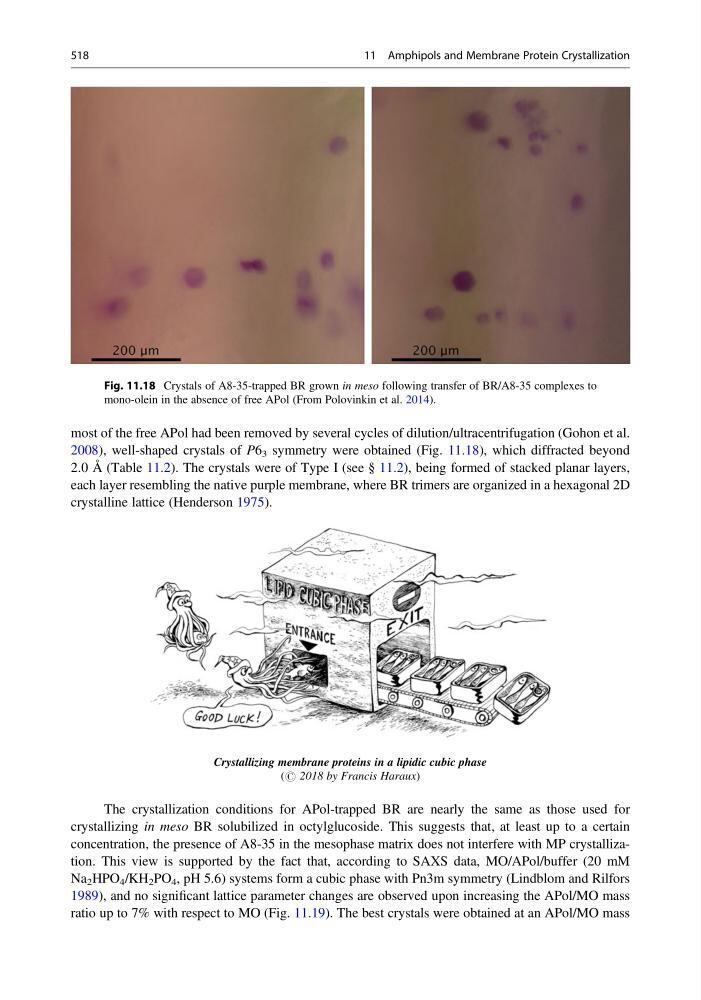
most of the free APol had been removed by several cycles of dilution/ultracentrifugation (Gohon et al.
2008), well-shaped crystals of P6 3 symmetry were obtained (Fig. 11.18), which diffracted beyond
2.0 Å (Table 11.2). The crystals were of Type I (see § 11.2), being formed of stacked planar layers,
each layer resembling the native purple membrane, where BR trimers are organized in a hexagonal 2D
crystalline lattice (Henderson 1975).
Crystallizing membrane proteins in a lipidic cubic phase
(# 2018 by Francis Haraux)
The crystallization conditions for APol-trapped BR are nearly the same as those used for
crystallizing in meso BR solubilized in octylglucoside. This suggests that, at least up to a certain
concentration, the presence of A8-35 in the mesophase matrix does not interfere with MP crystallization. This view is supported by the fact that, according to SAXS data, MO/APol/buffer (20 mM
Na 2 HPO 4 /KH 2 PO 4 , pH 5.6) systems form a cubic phase with Pn3m symmetry (Lindblom and Rilfors
1989), and no significant lattice parameter changes are observed upon increasing the APol/MO mass
ratio up to 7% with respect to MO (Fig. 11.19). The best crystals were obtained at an APol/MO mass
Fig. 11.18 Crystals of A8-35-trapped BR grown in meso following transfer of BR/A8-35 complexes to
mono-olein in the absence of free APol (From Polovinkin et al. 2014).
518
11 Amphipols and Membrane Protein Crystallization
2008), well-shaped crystals of P6 3 symmetry were obtained (Fig. 11.18), which diffracted beyond
2.0 Å (Table 11.2). The crystals were of Type I (see § 11.2), being formed of stacked planar layers,
each layer resembling the native purple membrane, where BR trimers are organized in a hexagonal 2D
crystalline lattice (Henderson 1975).
Crystallizing membrane proteins in a lipidic cubic phase
(# 2018 by Francis Haraux)
The crystallization conditions for APol-trapped BR are nearly the same as those used for
crystallizing in meso BR solubilized in octylglucoside. This suggests that, at least up to a certain
concentration, the presence of A8-35 in the mesophase matrix does not interfere with MP crystallization. This view is supported by the fact that, according to SAXS data, MO/APol/buffer (20 mM
Na 2 HPO 4 /KH 2 PO 4 , pH 5.6) systems form a cubic phase with Pn3m symmetry (Lindblom and Rilfors
1989), and no significant lattice parameter changes are observed upon increasing the APol/MO mass
ratio up to 7% with respect to MO (Fig. 11.19). The best crystals were obtained at an APol/MO mass
Fig. 11.18 Crystals of A8-35-trapped BR grown in meso following transfer of BR/A8-35 complexes to
mono-olein in the absence of free APol (From Polovinkin et al. 2014).
518
11 Amphipols and Membrane Protein Crystallization