

~22% (~107 kDa) for the bc 1 complex (486 kDa in its native dimeric form). It is surprising, however,
that the bc 1 dimer, with 26 TM α-helices, should bind about the same amount of A8-35 (~53 to 54 kDa)
as monomeric BR, which contains only seven helices + bound lipids (cf. Chap. 5, § 5.3.1.1). Given that
these measurements are difficult to carry out with good accuracy (if only because MPs tend to
oligomerize when separated from free APols, probably releasing APols; see Chap. 5), they clearly
have to be taken as provisional.
Functional measurements show that replacing the detergent belt with A8-35 does not affect the
electron transfer activity of the bc 1 complex (Charvolin et al. 2014).
Solution studies of bc 1 /A8-35 complexes showed them to be essentially monodisperse. Upon
SEC, they migrated as though they were slightly larger than bc 1 /detergent (DDM) ones. This behavior
is consistent with that observed for the calcium pump SERCA1a (A8-35 vs. DDM; Champeil et al.
2000), tOmpA (A8-35 vs. C 8 E 4 ; Zoonens et al. 2005, 2007), and OmpX (A8-35 vs. diC 6 PC; Catoire
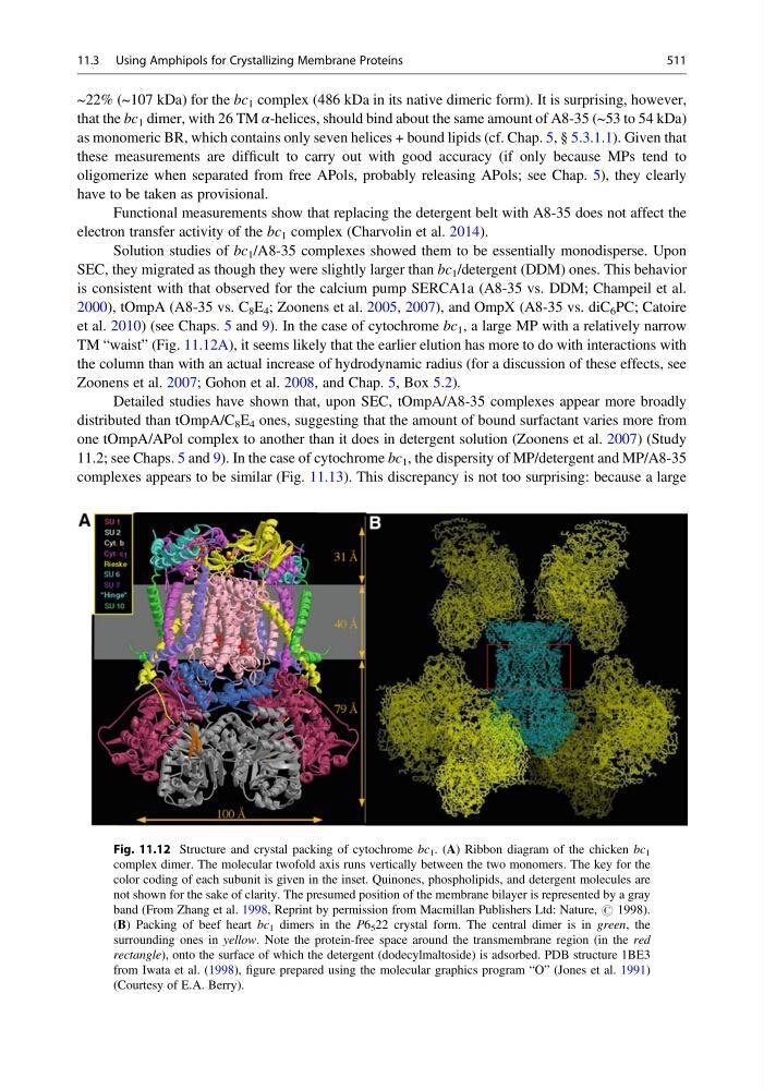
et al. 2010) (see Chaps. 5 and 9). In the case of cytochrome bc 1 , a large MP with a relatively narrow
TM “waist” (Fig. 11.12A), it seems likely that the earlier elution has more to do with interactions with
the column than with an actual increase of hydrodynamic radius (for a discussion of these effects, see
Zoonens et al. 2007; Gohon et al. 2008, and Chap. 5, Box 5.2).
Detailed studies have shown that, upon SEC, tOmpA/A8-35 complexes appear more broadly
distributed than tOmpA/C 8 E 4 ones, suggesting that the amount of bound surfactant varies more from
one tOmpA/APol complex to another than it does in detergent solution (Zoonens et al. 2007) (Study
11.2; see Chaps. 5 and 9). In the case of cytochrome bc 1 , the dispersity of MP/detergent and MP/A8-35
complexes appears to be similar (Fig. 11.13). This discrepancy is not too surprising: because a large
Fig. 11.12 Structure and crystal packing of cytochrome bc 1 . (A) Ribbon diagram of the chicken bc 1
complex dimer. The molecular twofold axis runs vertically between the two monomers. The key for the
color coding of each subunit is given in the inset. Quinones, phospholipids, and detergent molecules are
not shown for the sake of clarity. The presumed position of the membrane bilayer is represented by a gray
band (From Zhang et al. 1998, Reprint by permission from Macmillan Publishers Ltd: Nature, # 1998).
(B) Packing of beef heart bc 1 dimers in the P6 5 22 crystal form. The central dimer is in green, the
surrounding ones in yellow. Note the protein-free space around the transmembrane region (in the red
rectangle), onto the surface of which the detergent (dodecylmaltoside) is adsorbed. PDB structure 1BE3
from Iwata et al. (1998), figure prepared using the molecular graphics program “O” (Jones et al. 1991)
(Courtesy of E.A. Berry).
11.3 Using Amphipols for Crystallizing Membrane Proteins
511
that the bc 1 dimer, with 26 TM α-helices, should bind about the same amount of A8-35 (~53 to 54 kDa)
as monomeric BR, which contains only seven helices + bound lipids (cf. Chap. 5, § 5.3.1.1). Given that
these measurements are difficult to carry out with good accuracy (if only because MPs tend to
oligomerize when separated from free APols, probably releasing APols; see Chap. 5), they clearly
have to be taken as provisional.
Functional measurements show that replacing the detergent belt with A8-35 does not affect the
electron transfer activity of the bc 1 complex (Charvolin et al. 2014).
Solution studies of bc 1 /A8-35 complexes showed them to be essentially monodisperse. Upon
SEC, they migrated as though they were slightly larger than bc 1 /detergent (DDM) ones. This behavior
is consistent with that observed for the calcium pump SERCA1a (A8-35 vs. DDM; Champeil et al.
2000), tOmpA (A8-35 vs. C 8 E 4 ; Zoonens et al. 2005, 2007), and OmpX (A8-35 vs. diC 6 PC; Catoire
et al. 2010) (see Chaps. 5 and 9). In the case of cytochrome bc 1 , a large MP with a relatively narrow
TM “waist” (Fig. 11.12A), it seems likely that the earlier elution has more to do with interactions with
the column than with an actual increase of hydrodynamic radius (for a discussion of these effects, see
Zoonens et al. 2007; Gohon et al. 2008, and Chap. 5, Box 5.2).
Detailed studies have shown that, upon SEC, tOmpA/A8-35 complexes appear more broadly
distributed than tOmpA/C 8 E 4 ones, suggesting that the amount of bound surfactant varies more from
one tOmpA/APol complex to another than it does in detergent solution (Zoonens et al. 2007) (Study
11.2; see Chaps. 5 and 9). In the case of cytochrome bc 1 , the dispersity of MP/detergent and MP/A8-35
complexes appears to be similar (Fig. 11.13). This discrepancy is not too surprising: because a large
Fig. 11.12 Structure and crystal packing of cytochrome bc 1 . (A) Ribbon diagram of the chicken bc 1
complex dimer. The molecular twofold axis runs vertically between the two monomers. The key for the
color coding of each subunit is given in the inset. Quinones, phospholipids, and detergent molecules are
not shown for the sake of clarity. The presumed position of the membrane bilayer is represented by a gray
band (From Zhang et al. 1998, Reprint by permission from Macmillan Publishers Ltd: Nature, # 1998).
(B) Packing of beef heart bc 1 dimers in the P6 5 22 crystal form. The central dimer is in green, the
surrounding ones in yellow. Note the protein-free space around the transmembrane region (in the red
rectangle), onto the surface of which the detergent (dodecylmaltoside) is adsorbed. PDB structure 1BE3
from Iwata et al. (1998), figure prepared using the molecular graphics program “O” (Jones et al. 1991)
(Courtesy of E.A. Berry).
11.3 Using Amphipols for Crystallizing Membrane Proteins
511