

(Tribet et al. 1996; Gohon et al. 2008; Zoonens et al. 2014) (see Chap. 5). As observed for these
proteins, efficient retention into solution following dilution of the detergent under its CMC required
that the bc 1 be trapped with A8-35 at APol/protein mass ratios slightly higher than that eventually
measured in the final complexes. The mass ratio of bound APol, however, is much lower for
cytochrome bc 1 (~0.11 g bound A8-35 per g protein) than for BR (~2 g per g) (Gohon et al. 2008).
This is consistent with the different distribution of masses between TM and extramembrane regions: in
BR (27 kDa), the TM region represents ~75% of the mass of the protein, i.e. ~20 kDa, against only
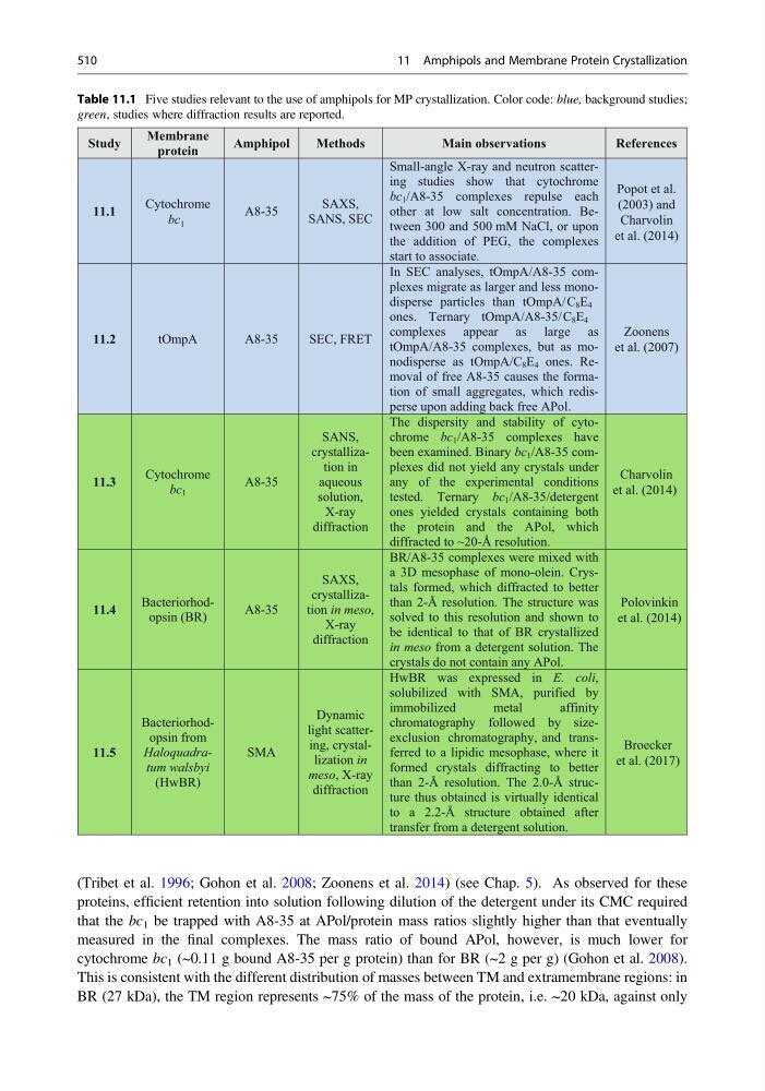
Table 11.1 Five studies relevant to the use of amphipols for MP crystallization. Color code: blue, background studies;
green, studies where diffraction results are reported.
Study
Membrane
protein
Amphipol
Methods
Main observations
References
11.1
Cytochrome
bc 1
A8-35
SAXS,
SANS, SEC
Small-angle X-ray and neutron scattering studies show that cytochrome
bc 1 /A8-35 complexes repulse each
other at low salt concentration. Between 300 and 500 mM NaCl, or upon
the addition of PEG, the complexes
start to associate.
11.2
tOmpA
A8-35
SEC, FRET
In SEC analyses, tOmpA/A8-35 complexes migrate as larger and less monodisperse particles than tOmpA/C 8 E 4
ones. Ternary tOmpA/A8-35/C 8 E 4
complexes appear as large as
tOmpA/A8-35 complexes, but as monodisperse as tOmpA/C 8 E 4 ones. Removal of free A8-35 causes the formation of small aggregates, which redisperse upon adding back free APol.
11.3
Cytochrome
bc 1
A8-35
SANS,
crystallization in
aqueous
solution,
X-ray
diffraction
The dispersity and stability of cytochrome bc 1 /A8-35 complexes have
been examined. Binary bc 1 /A8-35 complexes did not yield any crystals under
any of the experimental conditions
tested. Ternary bc 1 /A8-35/detergent
ones yielded crystals containing both
the protein and the APol, which
diffracted to ~20-Å resolution.
11.4
Bacteriorhodopsin (BR)
A8-35
SAXS,
crystallization in meso,
X-ray
diffraction
BR/A8-35 complexes were mixed with
a 3D mesophase of mono-olein. Crystals formed, which diffracted to better
than 2-Å resolution. The structure was
solved to this resolution and shown to
be identical to that of BR crystallized
in meso from a detergent solution. The
crystals do not contain any APol.
11.5
Bacteriorhodopsin from
Haloquadratum walsbyi
(HwBR)
SMA
Dynamic
light scattering, crystallization in
meso, X-ray
diffraction
HwBR was expressed in E. coli,
solubilized with SMA, purified by
immobilized
metal
affinity
chromatography followed by sizeexclusion chromatography, and transferred to a lipidic mesophase, where it
formed crystals diffracting to better
than 2-Å resolution. The 2.0-Å structure thus obtained is virtually identical
to a 2.2-Å structure obtained after
transfer from a detergent solution.
Popot et al.
(2003) and
Charvolin
et al. (2014)
Zoonens
et al. (2007)
Charvolin
et al. (2014)
Polovinkin
et al. (2014)
Broecker
et al. (2017)
510
11 Amphipols and Membrane Protein Crystallization
proteins, efficient retention into solution following dilution of the detergent under its CMC required
that the bc 1 be trapped with A8-35 at APol/protein mass ratios slightly higher than that eventually
measured in the final complexes. The mass ratio of bound APol, however, is much lower for
cytochrome bc 1 (~0.11 g bound A8-35 per g protein) than for BR (~2 g per g) (Gohon et al. 2008).
This is consistent with the different distribution of masses between TM and extramembrane regions: in
BR (27 kDa), the TM region represents ~75% of the mass of the protein, i.e. ~20 kDa, against only
Table 11.1 Five studies relevant to the use of amphipols for MP crystallization. Color code: blue, background studies;
green, studies where diffraction results are reported.
Study
Membrane
protein
Amphipol
Methods
Main observations
References
11.1
Cytochrome
bc 1
A8-35
SAXS,
SANS, SEC
Small-angle X-ray and neutron scattering studies show that cytochrome
bc 1 /A8-35 complexes repulse each
other at low salt concentration. Between 300 and 500 mM NaCl, or upon
the addition of PEG, the complexes
start to associate.
11.2
tOmpA
A8-35
SEC, FRET
In SEC analyses, tOmpA/A8-35 complexes migrate as larger and less monodisperse particles than tOmpA/C 8 E 4
ones. Ternary tOmpA/A8-35/C 8 E 4
complexes appear as large as
tOmpA/A8-35 complexes, but as monodisperse as tOmpA/C 8 E 4 ones. Removal of free A8-35 causes the formation of small aggregates, which redisperse upon adding back free APol.
11.3
Cytochrome
bc 1
A8-35
SANS,
crystallization in
aqueous
solution,
X-ray
diffraction
The dispersity and stability of cytochrome bc 1 /A8-35 complexes have
been examined. Binary bc 1 /A8-35 complexes did not yield any crystals under
any of the experimental conditions
tested. Ternary bc 1 /A8-35/detergent
ones yielded crystals containing both
the protein and the APol, which
diffracted to ~20-Å resolution.
11.4
Bacteriorhodopsin (BR)
A8-35
SAXS,
crystallization in meso,
X-ray
diffraction
BR/A8-35 complexes were mixed with
a 3D mesophase of mono-olein. Crystals formed, which diffracted to better
than 2-Å resolution. The structure was
solved to this resolution and shown to
be identical to that of BR crystallized
in meso from a detergent solution. The
crystals do not contain any APol.
11.5
Bacteriorhodopsin from
Haloquadratum walsbyi
(HwBR)
SMA
Dynamic
light scattering, crystallization in
meso, X-ray
diffraction
HwBR was expressed in E. coli,
solubilized with SMA, purified by
immobilized
metal
affinity
chromatography followed by sizeexclusion chromatography, and transferred to a lipidic mesophase, where it
formed crystals diffracting to better
than 2-Å resolution. The 2.0-Å structure thus obtained is virtually identical
to a 2.2-Å structure obtained after
transfer from a detergent solution.
Popot et al.
(2003) and
Charvolin
et al. (2014)
Zoonens
et al. (2007)
Charvolin
et al. (2014)
Polovinkin
et al. (2014)
Broecker
et al. (2017)
510
11 Amphipols and Membrane Protein Crystallization