

has yielded excellent crystals, from which the structure could be solved at high resolution (§ 11.3.2).
We will discuss successively approaches (i), (ii), and (iv), on the basis of data presented in the studies
listed in Table 11.1. The results of the crystallization trials of Study 11.3 will be presented first, and
their outcome discussed with the help of solution studies described in Studies 11.1–11.3. The most
recent and very encouraging results of Studies 11.4 and 11.5 will then be presented, before moving on
to § 11.3.3, which discusses challenges and perspectives.
11.3.1 Crystallization of Cytochrome bc 1 /A8-35 Complexes in Aqueous Solutions:
Failures and Partial Successes and the Possible Reasons Underlying Them
Study 11.3 reports the results of the first attempts at crystallizing MP/APol complexes (Charvolin et al.
2014). The model MP chosen is the cytochrome bc 1 complex extracted from beef heart mitochondria.
Cytochrome bc 1 presents numerous advantages for such a study: the complex is abundant, colored,
rugged, and relatively easy to crystallize from detergent solutions under a variety of conditions, and it
features extended extramembrane domains, which represent ~4/5 of its mass (Berry et al. 2000)
(Fig. 11.12A). As a consequence, and despite the large number of α-helices and cofactors that compose
its TM region, Type II crystals can form in which protein/protein contacts are solely established
between extramembrane surfaces, and there is no or very little interaction between surfactant layers
(Fig. 11.12B). In Study 11.3, the formation, composition, dispersity, and stability of bc 1 /A8-35
complexes have been examined, as well as their solution behavior as a function of such variables as
the presence of excess APol or the salt concentration. Crystallization attempts were carried out both of
binary bc 1 /A8-35 complexes and of ternary bc 1 /A8-35/detergent ones.
Complexation of cytochrome bc 1 by A8-35 took place efficiently under conditions similar to
those described for other MPs, e.g. the TM domain of OmpA (tOmpA) (Zoonens et al. 2005) or BR
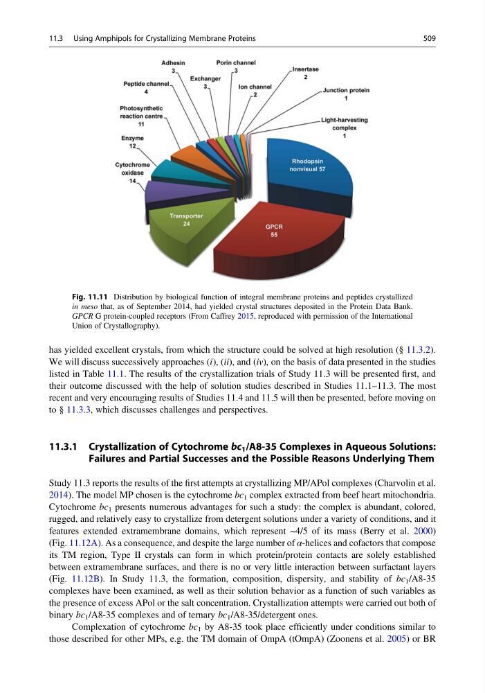
Fig. 11.11 Distribution by biological function of integral membrane proteins and peptides crystallized
in meso that, as of September 2014, had yielded crystal structures deposited in the Protein Data Bank.
GPCR G protein-coupled receptors (From Caffrey 2015, reproduced with permission of the International
Union of Crystallography).
11.3 Using Amphipols for Crystallizing Membrane Proteins
509
We will discuss successively approaches (i), (ii), and (iv), on the basis of data presented in the studies
listed in Table 11.1. The results of the crystallization trials of Study 11.3 will be presented first, and
their outcome discussed with the help of solution studies described in Studies 11.1–11.3. The most
recent and very encouraging results of Studies 11.4 and 11.5 will then be presented, before moving on
to § 11.3.3, which discusses challenges and perspectives.
11.3.1 Crystallization of Cytochrome bc 1 /A8-35 Complexes in Aqueous Solutions:
Failures and Partial Successes and the Possible Reasons Underlying Them
Study 11.3 reports the results of the first attempts at crystallizing MP/APol complexes (Charvolin et al.
2014). The model MP chosen is the cytochrome bc 1 complex extracted from beef heart mitochondria.
Cytochrome bc 1 presents numerous advantages for such a study: the complex is abundant, colored,
rugged, and relatively easy to crystallize from detergent solutions under a variety of conditions, and it
features extended extramembrane domains, which represent ~4/5 of its mass (Berry et al. 2000)
(Fig. 11.12A). As a consequence, and despite the large number of α-helices and cofactors that compose
its TM region, Type II crystals can form in which protein/protein contacts are solely established
between extramembrane surfaces, and there is no or very little interaction between surfactant layers
(Fig. 11.12B). In Study 11.3, the formation, composition, dispersity, and stability of bc 1 /A8-35
complexes have been examined, as well as their solution behavior as a function of such variables as
the presence of excess APol or the salt concentration. Crystallization attempts were carried out both of
binary bc 1 /A8-35 complexes and of ternary bc 1 /A8-35/detergent ones.
Complexation of cytochrome bc 1 by A8-35 took place efficiently under conditions similar to
those described for other MPs, e.g. the TM domain of OmpA (tOmpA) (Zoonens et al. 2005) or BR
Fig. 11.11 Distribution by biological function of integral membrane proteins and peptides crystallized
in meso that, as of September 2014, had yielded crystal structures deposited in the Protein Data Bank.
GPCR G protein-coupled receptors (From Caffrey 2015, reproduced with permission of the International
Union of Crystallography).
11.3 Using Amphipols for Crystallizing Membrane Proteins
509