

After relatively slow beginnings – due, in part, to practical difficulties in handling the samples, to
the initial absence of robots able to prepare them, and to the rather cumbersome recovery of the crystals
once they have formed – the approach has become strongly established as a major tool in MP
crystallogenesis (for reviews, see e.g. Rummel et al. 1998; Nollert 2004; Wiener 2004; Cherezov
et al. 2006; Hunte and Richers 2008; Caffrey 2009, 2011, 2015; Johansson et al. 2009; Cherezov 2011;
Ishchenko et al. 2014, 2017; Moraes et al. 2014; Bogorodskiy et al. 2015). As of September 2014, close
to 200 MPs, or about 10% of all published crystallographic MP structures, had been crystallized in meso
(Caffrey 2015). Their distribution by function is shown in Fig. 11.11. Success has been spectacular in
the field of GPCRs, whose crystallization from detergent solutions is notoriously difficult.
11.3 Using Amphipols for Crystallizing Membrane Proteins
As has been discussed in the case of NMR (cf. Chap. 10, Fig. 10.1), APols can be used upstream of
crystallization itself, so as to produce the target MPs, e.g. by folding them from inclusion bodies or by
expressing them in vitro. APol-trapped MPs thus obtained can be (i) used as such for crystallization
attempts; (ii) supplemented with a controlled amount of detergent so as to form ternary MP/APol/
detergent complexes; (iii) transferred to a detergent solution, from which to crystallize it using
conventional approaches; or (iv) transferred to a lipid mesophase. Approach (i) has been extensively
tried using the complexes formed between mitochondrial cytochrome bc 1 and A8-35, without ever
yielding any crystals, the possible reasons for which failure will be discussed below (§ 11.3.1).
Approach (ii) did yield crystals, although those have not been optimized yet (§ 11.3.1). Approach
(iii) has not yet been used, to our knowledge, and is essentially beyond the frame of this chapter, since
APols would be removed before starting crystallization attempts. Approach (iv) has been experimented
with, using BR from Halobacterium salinarum or from Haloquadratum walsbyi as the test protein, and
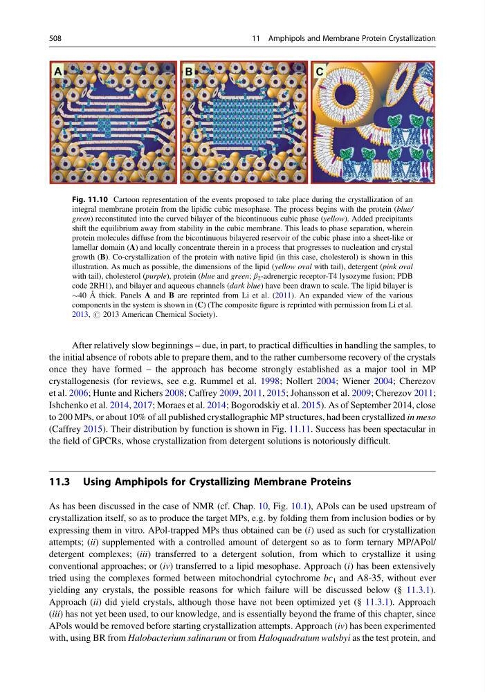
Fig. 11.10 Cartoon representation of the events proposed to take place during the crystallization of an
integral membrane protein from the lipidic cubic mesophase. The process begins with the protein (blue/
green) reconstituted into the curved bilayer of the bicontinuous cubic phase (yellow). Added precipitants
shift the equilibrium away from stability in the cubic membrane. This leads to phase separation, wherein
protein molecules diffuse from the bicontinuous bilayered reservoir of the cubic phase into a sheet-like or
lamellar domain (A) and locally concentrate therein in a process that progresses to nucleation and crystal
growth (B). Co-crystallization of the protein with native lipid (in this case, cholesterol) is shown in this
illustration. As much as possible, the dimensions of the lipid (yellow oval with tail), detergent (pink oval
with tail), cholesterol (purple), protein (blue and green; β 2 -adrenergic receptor-T4 lysozyme fusion; PDB
code 2RH1), and bilayer and aqueous channels (dark blue) have been drawn to scale. The lipid bilayer is
$40 Å thick. Panels A and B are reprinted from Li et al. (2011). An expanded view of the various
components in the system is shown in (C) (The composite figure is reprinted with permission from Li et al.
2013, # 2013 American Chemical Society).
508
11 Amphipols and Membrane Protein Crystallization
the initial absence of robots able to prepare them, and to the rather cumbersome recovery of the crystals
once they have formed – the approach has become strongly established as a major tool in MP
crystallogenesis (for reviews, see e.g. Rummel et al. 1998; Nollert 2004; Wiener 2004; Cherezov
et al. 2006; Hunte and Richers 2008; Caffrey 2009, 2011, 2015; Johansson et al. 2009; Cherezov 2011;
Ishchenko et al. 2014, 2017; Moraes et al. 2014; Bogorodskiy et al. 2015). As of September 2014, close
to 200 MPs, or about 10% of all published crystallographic MP structures, had been crystallized in meso
(Caffrey 2015). Their distribution by function is shown in Fig. 11.11. Success has been spectacular in
the field of GPCRs, whose crystallization from detergent solutions is notoriously difficult.
11.3 Using Amphipols for Crystallizing Membrane Proteins
As has been discussed in the case of NMR (cf. Chap. 10, Fig. 10.1), APols can be used upstream of
crystallization itself, so as to produce the target MPs, e.g. by folding them from inclusion bodies or by
expressing them in vitro. APol-trapped MPs thus obtained can be (i) used as such for crystallization
attempts; (ii) supplemented with a controlled amount of detergent so as to form ternary MP/APol/
detergent complexes; (iii) transferred to a detergent solution, from which to crystallize it using
conventional approaches; or (iv) transferred to a lipid mesophase. Approach (i) has been extensively
tried using the complexes formed between mitochondrial cytochrome bc 1 and A8-35, without ever
yielding any crystals, the possible reasons for which failure will be discussed below (§ 11.3.1).
Approach (ii) did yield crystals, although those have not been optimized yet (§ 11.3.1). Approach
(iii) has not yet been used, to our knowledge, and is essentially beyond the frame of this chapter, since
APols would be removed before starting crystallization attempts. Approach (iv) has been experimented
with, using BR from Halobacterium salinarum or from Haloquadratum walsbyi as the test protein, and
Fig. 11.10 Cartoon representation of the events proposed to take place during the crystallization of an
integral membrane protein from the lipidic cubic mesophase. The process begins with the protein (blue/
green) reconstituted into the curved bilayer of the bicontinuous cubic phase (yellow). Added precipitants
shift the equilibrium away from stability in the cubic membrane. This leads to phase separation, wherein
protein molecules diffuse from the bicontinuous bilayered reservoir of the cubic phase into a sheet-like or
lamellar domain (A) and locally concentrate therein in a process that progresses to nucleation and crystal
growth (B). Co-crystallization of the protein with native lipid (in this case, cholesterol) is shown in this
illustration. As much as possible, the dimensions of the lipid (yellow oval with tail), detergent (pink oval
with tail), cholesterol (purple), protein (blue and green; β 2 -adrenergic receptor-T4 lysozyme fusion; PDB
code 2RH1), and bilayer and aqueous channels (dark blue) have been drawn to scale. The lipid bilayer is
$40 Å thick. Panels A and B are reprinted from Li et al. (2011). An expanded view of the various
components in the system is shown in (C) (The composite figure is reprinted with permission from Li et al.
2013, # 2013 American Chemical Society).
508
11 Amphipols and Membrane Protein Crystallization