

been discussed in Chap. 2 (§ 2.4), tend to denature more rapidly in detergents with small
hydrophobic chains, which have a high CMC, than in those with long chains and low CMC.
The same search for minimal encumbrance by the surfactant belt has led to the use of small
amphiphilic additives like pentanetriol, which, by increasing the spontaneous curvature of the
surfactant/water interface, favor the formation of small micelles and can substitute for bulky
detergent molecules at points of close approach between surfactant belts (see Michel 1983;
Parker and Newstead 2012).
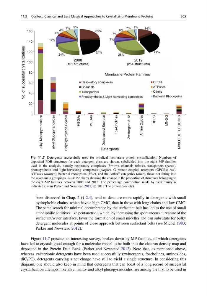
Figure 11.7 presents an interesting survey, broken down by MP families, of which detergents
have led to crystals good enough for a molecular model to be built into the electron density map and
deposited in the Protein Data Bank (Parker and Newstead 2012). Note that, as mentioned above,
whereas zwitterionic detergents have been used successfully (zwittergents, foscholines, aminoxides,
diC 7 PC), detergents carrying a net charge have still to yield a single structure. In considering this
diagram, one should also keep in mind that detergents that can boast of a long record of successful
crystallization attempts, like alkyl malto- and alkyl glucopyranosides, are among the first to be used in
0
alkyl Maltopyranosides
alkyl Glucopyranosides
Polyoxyethylene Glycols
HEGAs
MEGAs
Dodecanoyl Sucrose
Fos-Cholines
Zwittergent 3-12
Amine Oxides
MNG-3
DHPC
Detergents
No. of successful crystallizations
Respiratory complexes
24%
24%
29%
28%
7%
7%
7%
14%
2%
6%
24%
12%
4%
4%
7% 3%
Membrane Protein Families
2008
(121 structures)
2012
(254 structures)
GPCR
ATPases
Others
Bacterial Rhodopsins
Channels
Transporters
Photosynthetic & Light harvesting complexes
MIXED DETERGENTS
20
40
60
80
100
120
140
160
Fig. 11.7 Detergents successfully used for α-helical membrane protein crystallization. Numbers of
deposited PDB structures for each detergent class are shown, subdivided into the eight MP families
used in the analysis, namely respiratory complexes (brown), channels (black), transporters (green),
photosynthetic and light-harvesting complexes (purple), G protein-coupled receptors (GPCRs; red),
ATPases (orange), bacterial rhodopsins (blue), and the “other” categories (olive), those not fitting into
the seven main groupings. Inset: Pie charts showing the change in the proportion of structures belonging to
the eight MP families between 2008 and 2012. The percentage contribution made by each family is
indicated (From Parker and Newstead 2012, # 2012 The protein Society).
11.2 Context: Classical and Less Classical Approaches to Crystallizing Membrane Proteins
505
hydrophobic chains, which have a high CMC, than in those with long chains and low CMC.
The same search for minimal encumbrance by the surfactant belt has led to the use of small
amphiphilic additives like pentanetriol, which, by increasing the spontaneous curvature of the
surfactant/water interface, favor the formation of small micelles and can substitute for bulky
detergent molecules at points of close approach between surfactant belts (see Michel 1983;
Parker and Newstead 2012).
Figure 11.7 presents an interesting survey, broken down by MP families, of which detergents
have led to crystals good enough for a molecular model to be built into the electron density map and
deposited in the Protein Data Bank (Parker and Newstead 2012). Note that, as mentioned above,
whereas zwitterionic detergents have been used successfully (zwittergents, foscholines, aminoxides,
diC 7 PC), detergents carrying a net charge have still to yield a single structure. In considering this
diagram, one should also keep in mind that detergents that can boast of a long record of successful
crystallization attempts, like alkyl malto- and alkyl glucopyranosides, are among the first to be used in
0
alkyl Maltopyranosides
alkyl Glucopyranosides
Polyoxyethylene Glycols
HEGAs
MEGAs
Dodecanoyl Sucrose
Fos-Cholines
Zwittergent 3-12
Amine Oxides
MNG-3
DHPC
Detergents
No. of successful crystallizations
Respiratory complexes
24%
24%
29%
28%
7%
7%
7%
14%
2%
6%
24%
12%
4%
4%
7% 3%
Membrane Protein Families
2008
(121 structures)
2012
(254 structures)
GPCR
ATPases
Others
Bacterial Rhodopsins
Channels
Transporters
Photosynthetic & Light harvesting complexes
MIXED DETERGENTS
20
40
60
80
100
120
140
160
Fig. 11.7 Detergents successfully used for α-helical membrane protein crystallization. Numbers of
deposited PDB structures for each detergent class are shown, subdivided into the eight MP families
used in the analysis, namely respiratory complexes (brown), channels (black), transporters (green),
photosynthetic and light-harvesting complexes (purple), G protein-coupled receptors (GPCRs; red),
ATPases (orange), bacterial rhodopsins (blue), and the “other” categories (olive), those not fitting into
the seven main groupings. Inset: Pie charts showing the change in the proportion of structures belonging to
the eight MP families between 2008 and 2012. The percentage contribution made by each family is
indicated (From Parker and Newstead 2012, # 2012 The protein Society).
11.2 Context: Classical and Less Classical Approaches to Crystallizing Membrane Proteins
505