

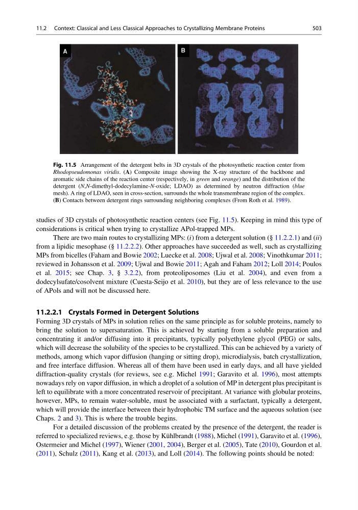
studies of 3D crystals of photosynthetic reaction centers (see Fig. 11.5). Keeping in mind this type of
considerations is critical when trying to crystallize APol-trapped MPs.
There are two main routes to crystallizing MPs: (i) from a detergent solution (§ 11.2.2.1) and (ii)
from a lipidic mesophase (§ 11.2.2.2). Other approaches have succeeded as well, such as crystallizing
MPs from bicelles (Faham and Bowie 2002; Luecke et al. 2008; Ujwal et al. 2008; Vinothkumar 2011;
reviewed in Johansson et al. 2009; Ujwal and Bowie 2011; Agah and Faham 2012; Loll 2014; Poulos
et al. 2015; see Chap. 3, § 3.2.2), from proteoliposomes (Liu et al. 2004), and even from a
dodecylsufate/cosolvent mixture (Cuesta-Seijo et al. 2010), but they are of less relevance to the use
of APols and will not be discussed here.
11.2.2.1 Crystals Formed in Detergent Solutions
Forming 3D crystals of MPs in solution relies on the same principle as for soluble proteins, namely to
bring the solution to supersaturation. This is achieved by starting from a soluble preparation and
concentrating it and/or diffusing into it precipitants, typically polyethylene glycol (PEG) or salts,
which will decrease the solubility of the species to be crystallized. This can be achieved by a variety of
methods, among which vapor diffusion (hanging or sitting drop), microdialysis, batch crystallization,
and free interface diffusion. Whereas all of them have been used in early days, and all have yielded
diffraction-quality crystals (for reviews, see e.g. Michel 1991; Garavito et al. 1996), most attempts
nowadays rely on vapor diffusion, in which a droplet of a solution of MP in detergent plus precipitant is
left to equilibrate with a more concentrated reservoir of precipitant. At variance with globular proteins,
however, MPs, to remain water-soluble, must be associated with a surfactant, typically a detergent,
which will provide the interface between their hydrophobic TM surface and the aqueous solution (see
Chaps. 2 and 3). This is where the trouble begins.
For a detailed discussion of the problems created by the presence of the detergent, the reader is
referred to specialized reviews, e.g. those by Kühlbrandt (1988), Michel (1991), Garavito et al. (1996),
Ostermeier and Michel (1997), Wiener (2001, 2004), Berger et al. (2005), Tate (2010), Gourdon et al.
(2011), Schulz (2011), Kang et al. (2013), and Loll (2014). The following points should be noted:
Fig. 11.5 Arrangement of the detergent belts in 3D crystals of the photosynthetic reaction center from
Rhodopseudomonas viridis. (A) Composite image showing the X-ray structure of the backbone and
aromatic side chains of the reaction center (respectively, in green and orange) and the distribution of the
detergent (N,N-dimethyl-dodecylamine-N-oxide; LDAO) as determined by neutron diffraction (blue
mesh). A ring of LDAO, seen in cross-section, surrounds the whole transmembrane region of the complex.
(B) Contacts between detergent rings surrounding neighboring complexes (From Roth et al. 1989).
11.2 Context: Classical and Less Classical Approaches to Crystallizing Membrane Proteins
503
considerations is critical when trying to crystallize APol-trapped MPs.
There are two main routes to crystallizing MPs: (i) from a detergent solution (§ 11.2.2.1) and (ii)
from a lipidic mesophase (§ 11.2.2.2). Other approaches have succeeded as well, such as crystallizing
MPs from bicelles (Faham and Bowie 2002; Luecke et al. 2008; Ujwal et al. 2008; Vinothkumar 2011;
reviewed in Johansson et al. 2009; Ujwal and Bowie 2011; Agah and Faham 2012; Loll 2014; Poulos
et al. 2015; see Chap. 3, § 3.2.2), from proteoliposomes (Liu et al. 2004), and even from a
dodecylsufate/cosolvent mixture (Cuesta-Seijo et al. 2010), but they are of less relevance to the use
of APols and will not be discussed here.
11.2.2.1 Crystals Formed in Detergent Solutions
Forming 3D crystals of MPs in solution relies on the same principle as for soluble proteins, namely to
bring the solution to supersaturation. This is achieved by starting from a soluble preparation and
concentrating it and/or diffusing into it precipitants, typically polyethylene glycol (PEG) or salts,
which will decrease the solubility of the species to be crystallized. This can be achieved by a variety of
methods, among which vapor diffusion (hanging or sitting drop), microdialysis, batch crystallization,
and free interface diffusion. Whereas all of them have been used in early days, and all have yielded
diffraction-quality crystals (for reviews, see e.g. Michel 1991; Garavito et al. 1996), most attempts
nowadays rely on vapor diffusion, in which a droplet of a solution of MP in detergent plus precipitant is
left to equilibrate with a more concentrated reservoir of precipitant. At variance with globular proteins,
however, MPs, to remain water-soluble, must be associated with a surfactant, typically a detergent,
which will provide the interface between their hydrophobic TM surface and the aqueous solution (see
Chaps. 2 and 3). This is where the trouble begins.
For a detailed discussion of the problems created by the presence of the detergent, the reader is
referred to specialized reviews, e.g. those by Kühlbrandt (1988), Michel (1991), Garavito et al. (1996),
Ostermeier and Michel (1997), Wiener (2001, 2004), Berger et al. (2005), Tate (2010), Gourdon et al.
(2011), Schulz (2011), Kang et al. (2013), and Loll (2014). The following points should be noted:
Fig. 11.5 Arrangement of the detergent belts in 3D crystals of the photosynthetic reaction center from
Rhodopseudomonas viridis. (A) Composite image showing the X-ray structure of the backbone and
aromatic side chains of the reaction center (respectively, in green and orange) and the distribution of the
detergent (N,N-dimethyl-dodecylamine-N-oxide; LDAO) as determined by neutron diffraction (blue
mesh). A ring of LDAO, seen in cross-section, surrounds the whole transmembrane region of the complex.
(B) Contacts between detergent rings surrounding neighboring complexes (From Roth et al. 1989).
11.2 Context: Classical and Less Classical Approaches to Crystallizing Membrane Proteins
503