

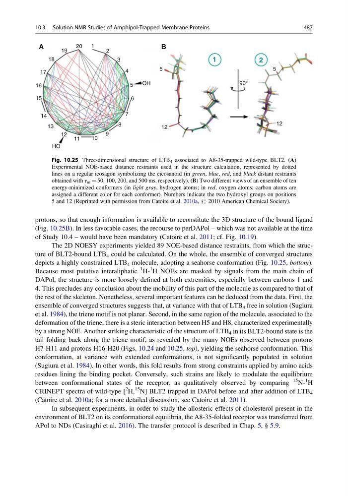
protons, so that enough information is available to reconstitute the 3D structure of the bound ligand
(Fig. 10.25B). In less favorable cases, the recourse to perDAPol – which was not available at the time
of Study 10.4 – would have been mandatory (Catoire et al. 2011; cf. Fig. 10.19).
The 2D NOESY experiments yielded 89 NOE-based distance restraints, from which the structure of BLT2-bound LTB 4 could be calculated. On the whole, the ensemble of converged structures
depicts a highly constrained LTB 4 molecule, adopting a seahorse conformation (Fig. 10.25, bottom).
Because most putative interaliphatic
1 H1 H NOEs are masked by signals from the main chain of
DAPol, the structure is more loosely defined at both extremities, especially between carbons 1 and
4. This precludes any conclusion about the mobility of this part of the molecule as compared to that of
the rest of the skeleton. Nonetheless, several important features can be deduced from the data. First, the
ensemble of converged structures suggests that, at variance with that of LTB 4 free in solution (Sugiura
et al. 1984), the triene motif is not planar. Second, in the same region of the molecule, associated to the
deformation of the triene, there is a steric interaction between H5 and H8, characterized experimentally
by a strong NOE. Another striking characteristic of the structure of LTB 4 in its BLT2-bound state is the
tail folding back along the triene motif, as revealed by the many NOEs observed between protons
H7-H11 and protons H16-H20 (Figs. 10.24 and 10.25, top), yielding the seahorse conformation. This
conformation, at variance with extended conformations, is not significantly populated in solution
(Sugiura et al. 1984). In other words, this fold results from strong constraints applied by amino acids
residues lining the binding pocket. Conversely, such strains are likely to modulate the equilibrium
between conformational states of the receptor, as qualitatively observed by comparing
15 N1 H
CRINEPT spectra of wild-type [
2 H,
15 N] BLT2 trapped in DAPol before and after addition of LTB 4
(Catoire et al. 2010a; for a more detailed discussion, see Catoire et al. 2011).
In subsequent experiments, in order to study the allosteric effects of cholesterol present in the
environment of BLT2 on its conformational equilibria, the A8-35-folded receptor was transferred from
APol to NDs (Casiraghi et al. 2016). The transfer protocol is described in Chap. 5, § 5.9.
A
20
1
2
3
4
5
6
7
8
9
10
11
12
13
14
15
16
17
18
19
OH
5
12
12
5
90°
1
2
HO
B
Fig. 10.25 Three-dimensional structure of LTB 4 associated to A8-35-trapped wild-type BLT2. (A)
Experimental NOE-based distance restraints used in the structure calculation, represented by dotted
lines on a regular icosagon symbolizing the eicosanoid (in green, blue, red, and black distant restraints
obtained with τ m ¼ 50, 100, 200, and 500 ms, respectively). (B) Two different views of an ensemble of ten
energy-minimized conformers (in light gray, hydrogen atoms; in red, oxygen atoms; carbon atoms are
assigned a different color for each conformer). Numbers indicate the two hydroxyl groups on positions
5 and 12 (Reprinted with permission from Catoire et al. 2010a, # 2010 American Chemical Society).
10.3 Solution NMR Studies of Amphipol-Trapped Membrane Proteins
487
(Fig. 10.25B). In less favorable cases, the recourse to perDAPol – which was not available at the time
of Study 10.4 – would have been mandatory (Catoire et al. 2011; cf. Fig. 10.19).
The 2D NOESY experiments yielded 89 NOE-based distance restraints, from which the structure of BLT2-bound LTB 4 could be calculated. On the whole, the ensemble of converged structures
depicts a highly constrained LTB 4 molecule, adopting a seahorse conformation (Fig. 10.25, bottom).
Because most putative interaliphatic
1 H1 H NOEs are masked by signals from the main chain of
DAPol, the structure is more loosely defined at both extremities, especially between carbons 1 and
4. This precludes any conclusion about the mobility of this part of the molecule as compared to that of
the rest of the skeleton. Nonetheless, several important features can be deduced from the data. First, the
ensemble of converged structures suggests that, at variance with that of LTB 4 free in solution (Sugiura
et al. 1984), the triene motif is not planar. Second, in the same region of the molecule, associated to the
deformation of the triene, there is a steric interaction between H5 and H8, characterized experimentally
by a strong NOE. Another striking characteristic of the structure of LTB 4 in its BLT2-bound state is the
tail folding back along the triene motif, as revealed by the many NOEs observed between protons
H7-H11 and protons H16-H20 (Figs. 10.24 and 10.25, top), yielding the seahorse conformation. This
conformation, at variance with extended conformations, is not significantly populated in solution
(Sugiura et al. 1984). In other words, this fold results from strong constraints applied by amino acids
residues lining the binding pocket. Conversely, such strains are likely to modulate the equilibrium
between conformational states of the receptor, as qualitatively observed by comparing
15 N1 H
CRINEPT spectra of wild-type [
2 H,
15 N] BLT2 trapped in DAPol before and after addition of LTB 4
(Catoire et al. 2010a; for a more detailed discussion, see Catoire et al. 2011).
In subsequent experiments, in order to study the allosteric effects of cholesterol present in the
environment of BLT2 on its conformational equilibria, the A8-35-folded receptor was transferred from
APol to NDs (Casiraghi et al. 2016). The transfer protocol is described in Chap. 5, § 5.9.
A
20
1
2
3
4
5
6
7
8
9
10
11
12
13
14
15
16
17
18
19
OH
5
12
12
5
90°
1
2
HO
B
Fig. 10.25 Three-dimensional structure of LTB 4 associated to A8-35-trapped wild-type BLT2. (A)
Experimental NOE-based distance restraints used in the structure calculation, represented by dotted
lines on a regular icosagon symbolizing the eicosanoid (in green, blue, red, and black distant restraints
obtained with τ m ¼ 50, 100, 200, and 500 ms, respectively). (B) Two different views of an ensemble of ten
energy-minimized conformers (in light gray, hydrogen atoms; in red, oxygen atoms; carbon atoms are
assigned a different color for each conformer). Numbers indicate the two hydroxyl groups on positions
5 and 12 (Reprinted with permission from Catoire et al. 2010a, # 2010 American Chemical Society).
10.3 Solution NMR Studies of Amphipol-Trapped Membrane Proteins
487