

DAPol was obtained by grafting perdeuterated octyl and isopropyl chains onto hydrogenated
polyacrylate, using the standard protocol used for synthesizing unlabeled A8-35 (HAPol; Gohon et al.
2004, 2006; Chap. 4). As the starting polymer is the same, the chain length distribution is also the same
and the only chemical difference between HAPol and DAPol comes from the slight batch-to-batch
variations of grafting ratios that are inherent to the synthesis protocol. As described in Chap. 4, the
physical and biochemical properties of HAPol and DAPol are indeed indistinguishable as long as they
are not affected by the isotopic labeling. The size, mass, solution behavior, SANS contrast match point,
etc. of free DAPol particles and individual molecules have been thoroughly studied by SEC, DLS,
SANS, AUC, and MS (see Chap. 4).
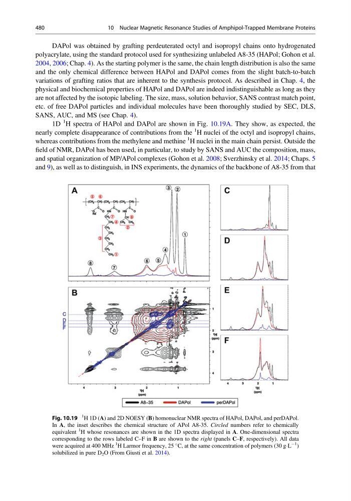
1D
1 H spectra of HAPol and DAPol are shown in Fig. 10.19A. They show, as expected, the
nearly complete disappearance of contributions from the
1 H nuclei of the octyl and isopropyl chains,
whereas contributions from the methylene and methine
1 H nuclei in the main chain persist. Outside the
field of NMR, DAPol has been used, in particular, to study by SANS and AUC the composition, mass,
and spatial organization of MP/APol complexes (Gohon et al. 2008; Sverzhinsky et al. 2014; Chaps. 5
and 9), as well as to distinguish, in INS experiments, the dynamics of the backbone of A8-35 from that
Fig. 10.19
1 H 1D (A) and 2D NOESY (B) homonuclear NMR spectra of HAPol, DAPol, and perDAPol.
In A, the inset describes the chemical structure of APol A8-35. Circled numbers refer to chemically
equivalent
1
H whose resonances are shown in the 1D spectra displayed in A. One-dimensional spectra
corresponding to the rows labeled C–F in B are shown to the right (panels C–F, respectively). All data
were acquired at 400 MHz
1
H Larmor frequency, 25
C, at the same concentration of polymers (30 gÁL
À1
)
solubilized in pure D 2 O (From Giusti et al. 2014).
480
10 Nuclear Magnetic Resonance Studies of Amphipol-Trapped Membrane Proteins
polyacrylate, using the standard protocol used for synthesizing unlabeled A8-35 (HAPol; Gohon et al.
2004, 2006; Chap. 4). As the starting polymer is the same, the chain length distribution is also the same
and the only chemical difference between HAPol and DAPol comes from the slight batch-to-batch
variations of grafting ratios that are inherent to the synthesis protocol. As described in Chap. 4, the
physical and biochemical properties of HAPol and DAPol are indeed indistinguishable as long as they
are not affected by the isotopic labeling. The size, mass, solution behavior, SANS contrast match point,
etc. of free DAPol particles and individual molecules have been thoroughly studied by SEC, DLS,
SANS, AUC, and MS (see Chap. 4).
1D
1 H spectra of HAPol and DAPol are shown in Fig. 10.19A. They show, as expected, the
nearly complete disappearance of contributions from the
1 H nuclei of the octyl and isopropyl chains,
whereas contributions from the methylene and methine
1 H nuclei in the main chain persist. Outside the
field of NMR, DAPol has been used, in particular, to study by SANS and AUC the composition, mass,
and spatial organization of MP/APol complexes (Gohon et al. 2008; Sverzhinsky et al. 2014; Chaps. 5
and 9), as well as to distinguish, in INS experiments, the dynamics of the backbone of A8-35 from that
Fig. 10.19
1 H 1D (A) and 2D NOESY (B) homonuclear NMR spectra of HAPol, DAPol, and perDAPol.
In A, the inset describes the chemical structure of APol A8-35. Circled numbers refer to chemically
equivalent
1
H whose resonances are shown in the 1D spectra displayed in A. One-dimensional spectra
corresponding to the rows labeled C–F in B are shown to the right (panels C–F, respectively). All data
were acquired at 400 MHz
1
H Larmor frequency, 25
C, at the same concentration of polymers (30 gÁL
À1
)
solubilized in pure D 2 O (From Giusti et al. 2014).
480
10 Nuclear Magnetic Resonance Studies of Amphipol-Trapped Membrane Proteins