

of its side chains (Tehei et al. 2014; Chap. 4). In sNMR, it has been used, as described above, to
improve spectra and to map MP/APol contacts (Zoonens et al. 2005; Catoire et al. 2010b; Planchard
et al. 2014). Its use has been critical in experiments involving the measurement of heteronuclear
(Catoire et al. 2009) and homonuclear (Catoire et al. 2010a) NOE signals.
Because contributions from the
1 H nuclei in the main chain remain a hindrance both in INS
studies of the dynamics of APol-trapped MPs and in NMR experiments involving the use of
1 H1 H or
1 H13 C NOE signals, a perdeuterated version of A8-35 (perDAPol) has been developed (Fig. 10.6C).
From the point of view of chemical synthesis, this was more of a challenge, because it necessitated the
synthesis of a perdeuterated polyacrylate with the same average length and dispersity as the commercial
hydrogenated product used for the syntheses of HAPol and DAPol (see Chap. 4). Once obtained, the
perdeuterated polyacrylate was grafted with perdeuterated octylamine and isopropylamine according to
the regular procedure for synthesizing HAPol and DAPol (Giusti et al. 2014).
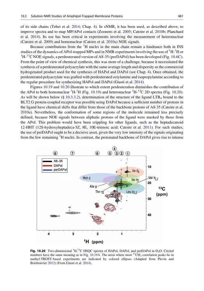
Figures 10.19 and 10.20 illustrate to which extent perdeuteration diminishes the contribution of
the APol to both homonuclear
1 H1 H (Fig. 10.19) and heteronuclear
1 H13 C 2D spectra (Fig. 10.20).
As will be shown below (§ 10.3.3.2), determination of the structure of the ligand LTB 4 bound to the
BLT2 G protein-coupled receptor was possible using DAPol because a sufficient number of protons in
the ligand have chemical shifts that differ from those of the backbone protons of A8-35 (Catoire et al.
2010a). Nevertheless, the conformation of some regions of the molecule remained less precisely
defined, because NOE signals between aliphatic protons of the ligand were masked by those from
the APol. This problem would have been crippling for other ligands, such as the heptadecanoid
12-HHT (12S-hydroxyheptadeca-5Z, 8E, 10E-trienoic acid; Catoire et al. 2011). For such studies,
the use of perDAPol ought to be a decisive asset, given the very low intensity of the signals originating
from the few remaining
1 H nuclei. In contrast, the protonated backbone of DAPol gives rise to intense
Fig. 10.20 Two-dimensional
1
H,
13
C HSQC spectra of HAPol, DAPol, and perDAPol in D 2 O. Circled
numbers have the same meaning as in Fig. 10.19A. The areas where most
13
CH 3 correlation peaks lie in
methyl-TROSY-based experiments are indicated by colored ellipses (Adapted from Plevin and
Boisbouvier 2012) (From Giusti et al. 2014).
10.3 Solution NMR Studies of Amphipol-Trapped Membrane Proteins
481
improve spectra and to map MP/APol contacts (Zoonens et al. 2005; Catoire et al. 2010b; Planchard
et al. 2014). Its use has been critical in experiments involving the measurement of heteronuclear
(Catoire et al. 2009) and homonuclear (Catoire et al. 2010a) NOE signals.
Because contributions from the
1 H nuclei in the main chain remain a hindrance both in INS
studies of the dynamics of APol-trapped MPs and in NMR experiments involving the use of
1 H1 H or
1 H13 C NOE signals, a perdeuterated version of A8-35 (perDAPol) has been developed (Fig. 10.6C).
From the point of view of chemical synthesis, this was more of a challenge, because it necessitated the
synthesis of a perdeuterated polyacrylate with the same average length and dispersity as the commercial
hydrogenated product used for the syntheses of HAPol and DAPol (see Chap. 4). Once obtained, the
perdeuterated polyacrylate was grafted with perdeuterated octylamine and isopropylamine according to
the regular procedure for synthesizing HAPol and DAPol (Giusti et al. 2014).
Figures 10.19 and 10.20 illustrate to which extent perdeuteration diminishes the contribution of
the APol to both homonuclear
1 H1 H (Fig. 10.19) and heteronuclear
1 H13 C 2D spectra (Fig. 10.20).
As will be shown below (§ 10.3.3.2), determination of the structure of the ligand LTB 4 bound to the
BLT2 G protein-coupled receptor was possible using DAPol because a sufficient number of protons in
the ligand have chemical shifts that differ from those of the backbone protons of A8-35 (Catoire et al.
2010a). Nevertheless, the conformation of some regions of the molecule remained less precisely
defined, because NOE signals between aliphatic protons of the ligand were masked by those from
the APol. This problem would have been crippling for other ligands, such as the heptadecanoid
12-HHT (12S-hydroxyheptadeca-5Z, 8E, 10E-trienoic acid; Catoire et al. 2011). For such studies,
the use of perDAPol ought to be a decisive asset, given the very low intensity of the signals originating
from the few remaining
1 H nuclei. In contrast, the protonated backbone of DAPol gives rise to intense
Fig. 10.20 Two-dimensional
1
H,
13
C HSQC spectra of HAPol, DAPol, and perDAPol in D 2 O. Circled
numbers have the same meaning as in Fig. 10.19A. The areas where most
13
CH 3 correlation peaks lie in
methyl-TROSY-based experiments are indicated by colored ellipses (Adapted from Plevin and
Boisbouvier 2012) (From Giusti et al. 2014).
10.3 Solution NMR Studies of Amphipol-Trapped Membrane Proteins
481