

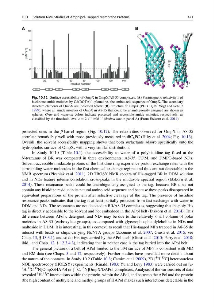
protected ones in the β-barrel region (Fig. 10.12). The relaxivities observed for OmpX in A8-35
correlate remarkably well with those previously measured in diC 6 PC (Hilty et al. 2004; Fig. 10.13).
Overall, the solvent accessibility mapping shows that both surfactants adsorb specifically onto the
hydrophobic surface of OmpX, with a very similar distribution.
In Study 10.10 (Table 10.1), the accessibility to water of a polyhistidine tag fused at the
N-terminus of BR was compared in three environments, A8-35, DDM, and DMPC-based NDs.
Solvent-accessible imidazole protons of the histidine ring experience proton exchange rates with the
surrounding water molecules in the fast chemical exchange regime and thus are not detectable in the
NMR spectrum (Plesniak et al. 2011). 2D TROSY NMR spectra of His-tagged BR in DDM solution
and in NDs feature intense correlation cross-peaks in the imidazole spectral region (Etzkorn et al.
2014). These resonance peaks could be unambiguously assigned to the tag, because BR does not
contain any histidine residue in its natural amino acid sequence and because these peaks disappeared in
equivalent preparations of the protein after selective cleavage of the tag. The presence of histidine
resonance peaks indicates that the tag is at least partially protected from fast exchange with water in
DDM and NDs. The resonances are not detected in BR/A8-35 complexes, suggesting that the poly-His
tag is directly accessible to the solvent and not embedded in the APol belt (Etzkorn et al. 2014). This
difference between APols, detergent, and NDs may be due to the relatively small volume of polar
moieties in A8-35 (carboxylate groups), as compared with glycerophosphatidylcholine in NDs and
maltoside in DDM. It is interesting, in this context, to recall that His-tagged MPs trapped in A8-35 do
interact with beads or chips carrying Ni/NTA groups (Zoonens et al. 2007; Giusti et al. 2015; see
Chap. 13, § 13.3.1), and so do His-tags carried by the APol itself (Giusti et al. 2015; Perry et al. 2018;
ibid., and Chap. 12, § 12.3.4.3), indicating that in neither case is the tag buried into the APol belt.
The general picture of a belt of APol limited to the TM surface of MPs is consistent with MD
and EM data (see Chaps. 5 and 12, respectively). Further studies have provided more details about
the nature of the contacts. In Study 10.2 (Table 10.3; Catoire et al. 2009), 2D [
1 H,
13 C] heteronuclear
NOE spectroscopy (HOESY) experiments (Rinaldi 1983; Yu and Levy 1983) were carried out on [u2 H,
13 C,
15 N]OmpX/HAPol or [
13 C,
15 N]OmpX/DAPol complexes. Analysis of the various sets of data
revealed
1 H13 C interactions within the protein, within the APol, and between the APol and the protein
(the high content of methylene and methyl groups of HAPol makes such interactions detectable in the
Fig. 10.12 Surface accessibility of OmpX in OmpX/A8-35 complexes. (A) Paramagnetic relaxivity ε of
backbone amide moieties by Gd(DOTA)
À , plotted vs. the amino acid sequence of OmpX. The secondary
structure elements of OmpX are indicated below. (B) Structure of OmpX (PDB 1QJ8; Vogt and Schulz
1999), where all amide moieties of OmpX in A8-35 that could be unambiguously assigned are shown as
spheres. Gray and magenta colors indicate protected and accessible amide moieties, respectively, as
classified by the threshold level ε ¼ 2 s
À1
ÁmM
À1 (dashed line in panel A) (From Etzkorn et al. 2014).
10.3 Solution NMR Studies of Amphipol-Trapped Membrane Proteins
471
correlate remarkably well with those previously measured in diC 6 PC (Hilty et al. 2004; Fig. 10.13).
Overall, the solvent accessibility mapping shows that both surfactants adsorb specifically onto the
hydrophobic surface of OmpX, with a very similar distribution.
In Study 10.10 (Table 10.1), the accessibility to water of a polyhistidine tag fused at the
N-terminus of BR was compared in three environments, A8-35, DDM, and DMPC-based NDs.
Solvent-accessible imidazole protons of the histidine ring experience proton exchange rates with the
surrounding water molecules in the fast chemical exchange regime and thus are not detectable in the
NMR spectrum (Plesniak et al. 2011). 2D TROSY NMR spectra of His-tagged BR in DDM solution
and in NDs feature intense correlation cross-peaks in the imidazole spectral region (Etzkorn et al.
2014). These resonance peaks could be unambiguously assigned to the tag, because BR does not
contain any histidine residue in its natural amino acid sequence and because these peaks disappeared in
equivalent preparations of the protein after selective cleavage of the tag. The presence of histidine
resonance peaks indicates that the tag is at least partially protected from fast exchange with water in
DDM and NDs. The resonances are not detected in BR/A8-35 complexes, suggesting that the poly-His
tag is directly accessible to the solvent and not embedded in the APol belt (Etzkorn et al. 2014). This
difference between APols, detergent, and NDs may be due to the relatively small volume of polar
moieties in A8-35 (carboxylate groups), as compared with glycerophosphatidylcholine in NDs and
maltoside in DDM. It is interesting, in this context, to recall that His-tagged MPs trapped in A8-35 do
interact with beads or chips carrying Ni/NTA groups (Zoonens et al. 2007; Giusti et al. 2015; see
Chap. 13, § 13.3.1), and so do His-tags carried by the APol itself (Giusti et al. 2015; Perry et al. 2018;
ibid., and Chap. 12, § 12.3.4.3), indicating that in neither case is the tag buried into the APol belt.
The general picture of a belt of APol limited to the TM surface of MPs is consistent with MD
and EM data (see Chaps. 5 and 12, respectively). Further studies have provided more details about
the nature of the contacts. In Study 10.2 (Table 10.3; Catoire et al. 2009), 2D [
1 H,
13 C] heteronuclear
NOE spectroscopy (HOESY) experiments (Rinaldi 1983; Yu and Levy 1983) were carried out on [u2 H,
13 C,
15 N]OmpX/HAPol or [
13 C,
15 N]OmpX/DAPol complexes. Analysis of the various sets of data
revealed
1 H13 C interactions within the protein, within the APol, and between the APol and the protein
(the high content of methylene and methyl groups of HAPol makes such interactions detectable in the
Fig. 10.12 Surface accessibility of OmpX in OmpX/A8-35 complexes. (A) Paramagnetic relaxivity ε of
backbone amide moieties by Gd(DOTA)
À , plotted vs. the amino acid sequence of OmpX. The secondary
structure elements of OmpX are indicated below. (B) Structure of OmpX (PDB 1QJ8; Vogt and Schulz
1999), where all amide moieties of OmpX in A8-35 that could be unambiguously assigned are shown as
spheres. Gray and magenta colors indicate protected and accessible amide moieties, respectively, as
classified by the threshold level ε ¼ 2 s
À1
ÁmM
À1 (dashed line in panel A) (From Etzkorn et al. 2014).
10.3 Solution NMR Studies of Amphipol-Trapped Membrane Proteins
471