

with properties identical to those formed by the native protein (Pocanschi et al. 2006) (see Chap. 6,
§ 6.3.1.2). There is therefore no doubt that the structures of APol-trapped tOmpA and OmpX are
extremely close if not identical to those of the proteins solubilized in detergent solutions that have been
used in X-ray and earlier sNMR studies.
Early works aimed at investigating to which extent sNMR of MP/APol complexes was practical.
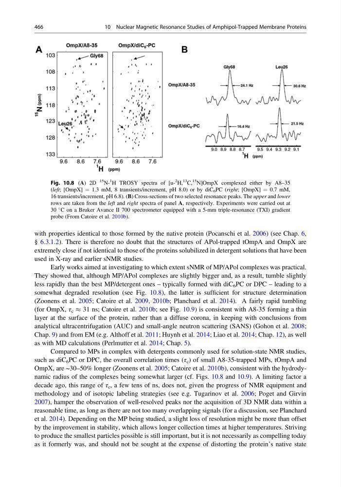
They showed that, although MP/APol complexes are slightly bigger and, as a result, tumble slightly
less rapidly than the best MP/detergent ones – typically formed with diC 6 PC or DPC – leading to a
somewhat degraded resolution (see Fig. 10.8), the latter is sufficient for structure determination
(Zoonens et al. 2005; Catoire et al. 2009, 2010b; Planchard et al. 2014). A fairly rapid tumbling
(for OmpX, τ c % 31 ns; Catoire et al. 2010b; see Fig. 10.9) is consistent with A8-35 forming a thin
layer at the surface of the protein, rather than a diffuse corona, in keeping with conclusions from
analytical ultracentrifugation (AUC) and small-angle neutron scattering (SANS) (Gohon et al. 2008;
Chap. 9) and from EM (e.g. Althoff et al. 2011; Huynh et al. 2014; Liao et al. 2014; Chap. 12), as well
as with MD calculations (Perlmutter et al. 2014; Chap. 5).
Compared to MPs in complex with detergents commonly used for solution-state NMR studies,
such as diC 6 PC or DPC, the overall correlation times (τ c ) of small A8-35-trapped MPs, tOmpA and
OmpX, are ~30–50% longer (Zoonens et al. 2005; Catoire et al. 2010b), consistent with the hydrodynamic radius of the complexes being somewhat larger (cf. Figs. 10.8 and 10.9). A limiting factor a
decade ago, this range of τ c , a few tens of ns, does not, given the progress of NMR equipment and
methodology and of isotopic labeling strategies (see e.g. Tugarinov et al. 2006; Poget and Girvin
2007), hamper the observation of well-resolved peaks nor the acquisition of 3D NMR data within a
reasonable time, as long as there are not too many overlapping signals (for a discussion, see Planchard
et al. 2014). Depending on the MP being studied, a slight loss of resolution might be more than offset
by the improvement in stability, which allows longer collection times at higher temperatures. Striving
to produce the smallest particles possible is still important, but it is not necessarily as compelling today
as it formerly was, and should not be sought at the expense of distorting the protein’s native state
Fig. 10.8 (A) 2D
15
N1
H TROSY spectra of [u2
H,
13 C,
15 N]OmpX complexed either by A8–35
(left; [OmpX] ¼ 1.3 mM, 8 transients/increment, pH 8.0) or by diC 6 PC (right; [OmpX] ¼ 0.7 mM,
16 transients/increment, pH 6.8). (B) Cross-sections of two selected resonance peaks. The upper and lower
rows are taken from the left and right spectra of panel A, respectively. Experiments were carried out at
30
C on a Bruker Avance II 700 spectrometer equipped with a 5-mm triple-resonance (TXI) gradient
probe (From Catoire et al. 2010b).
466
10 Nuclear Magnetic Resonance Studies of Amphipol-Trapped Membrane Proteins
§ 6.3.1.2). There is therefore no doubt that the structures of APol-trapped tOmpA and OmpX are
extremely close if not identical to those of the proteins solubilized in detergent solutions that have been
used in X-ray and earlier sNMR studies.
Early works aimed at investigating to which extent sNMR of MP/APol complexes was practical.
They showed that, although MP/APol complexes are slightly bigger and, as a result, tumble slightly
less rapidly than the best MP/detergent ones – typically formed with diC 6 PC or DPC – leading to a
somewhat degraded resolution (see Fig. 10.8), the latter is sufficient for structure determination
(Zoonens et al. 2005; Catoire et al. 2009, 2010b; Planchard et al. 2014). A fairly rapid tumbling
(for OmpX, τ c % 31 ns; Catoire et al. 2010b; see Fig. 10.9) is consistent with A8-35 forming a thin
layer at the surface of the protein, rather than a diffuse corona, in keeping with conclusions from
analytical ultracentrifugation (AUC) and small-angle neutron scattering (SANS) (Gohon et al. 2008;
Chap. 9) and from EM (e.g. Althoff et al. 2011; Huynh et al. 2014; Liao et al. 2014; Chap. 12), as well
as with MD calculations (Perlmutter et al. 2014; Chap. 5).
Compared to MPs in complex with detergents commonly used for solution-state NMR studies,
such as diC 6 PC or DPC, the overall correlation times (τ c ) of small A8-35-trapped MPs, tOmpA and
OmpX, are ~30–50% longer (Zoonens et al. 2005; Catoire et al. 2010b), consistent with the hydrodynamic radius of the complexes being somewhat larger (cf. Figs. 10.8 and 10.9). A limiting factor a
decade ago, this range of τ c , a few tens of ns, does not, given the progress of NMR equipment and
methodology and of isotopic labeling strategies (see e.g. Tugarinov et al. 2006; Poget and Girvin
2007), hamper the observation of well-resolved peaks nor the acquisition of 3D NMR data within a
reasonable time, as long as there are not too many overlapping signals (for a discussion, see Planchard
et al. 2014). Depending on the MP being studied, a slight loss of resolution might be more than offset
by the improvement in stability, which allows longer collection times at higher temperatures. Striving
to produce the smallest particles possible is still important, but it is not necessarily as compelling today
as it formerly was, and should not be sought at the expense of distorting the protein’s native state
Fig. 10.8 (A) 2D
15
N1
H TROSY spectra of [u2
H,
13 C,
15 N]OmpX complexed either by A8–35
(left; [OmpX] ¼ 1.3 mM, 8 transients/increment, pH 8.0) or by diC 6 PC (right; [OmpX] ¼ 0.7 mM,
16 transients/increment, pH 6.8). (B) Cross-sections of two selected resonance peaks. The upper and lower
rows are taken from the left and right spectra of panel A, respectively. Experiments were carried out at
30
C on a Bruker Avance II 700 spectrometer equipped with a 5-mm triple-resonance (TXI) gradient
probe (From Catoire et al. 2010b).
466
10 Nuclear Magnetic Resonance Studies of Amphipol-Trapped Membrane Proteins