

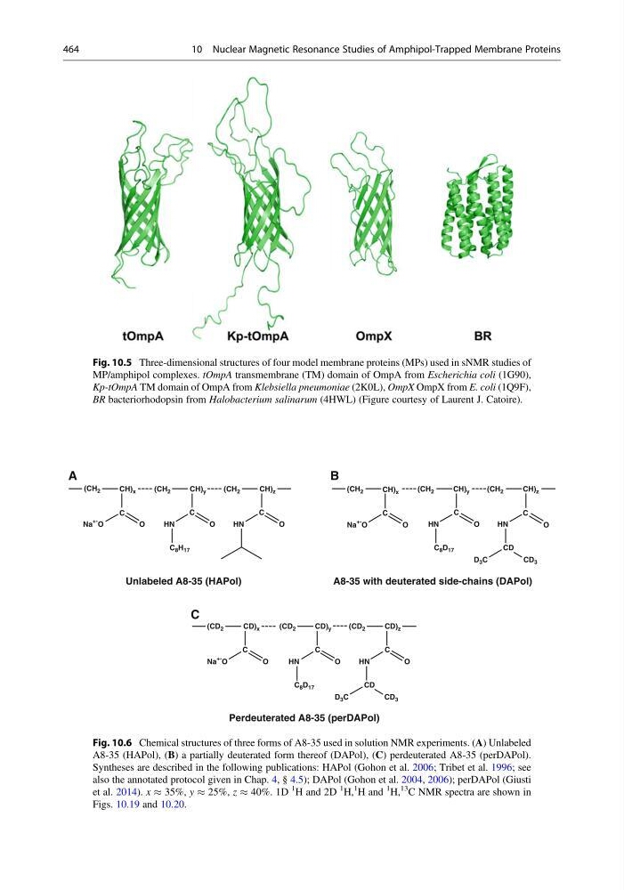
Fig. 10.5 Three-dimensional structures of four model membrane proteins (MPs) used in sNMR studies of
MP/amphipol complexes. tOmpA transmembrane (TM) domain of OmpA from Escherichia coli (1G90),
Kp-tOmpA TM domain of OmpA from Klebsiella pneumoniae (2K0L), OmpX OmpX from E. coli (1Q9F),
BR bacteriorhodopsin from Halobacterium salinarum (4HWL) (Figure courtesy of Laurent J. Catoire).
(CH 2
(CH 2
(CH 2
CH) x
CH) y
CH) z
(CH 2
(CH 2
(CH 2
CH) x
CH) y
CH) z
(CD 2
(CD 2
(CD 2
CD) x
CD) y
CD) z
HN
HN
C 8 H 17
Unlabeled A8-35 (HAPol)
A8-35 with deuterated side-chains (DAPol)
A
B
C
Perdeuterated A8-35 (perDAPol)
Na
+- O
Na
+- O
Na
+O
O
C
O
C
O
O
O
C
C
C
C
C
O
O
H N
H N
HN
HN
C 8 D 17
C 8 D 17
CD 3
D 3 C
CD
CD 3
D 3 C
CD
C
C
O
O
Fig. 10.6 Chemical structures of three forms of A8-35 used in solution NMR experiments. (A) Unlabeled
A8-35 (HAPol), (B) a partially deuterated form thereof (DAPol), (C) perdeuterated A8-35 (perDAPol).
Syntheses are described in the following publications: HAPol (Gohon et al. 2006; Tribet et al. 1996; see
also the annotated protocol given in Chap. 4, § 4.5); DAPol (Gohon et al. 2004, 2006); perDAPol (Giusti
et al. 2014). x % 35%, y % 25%, z % 40%. 1D
1
H and 2D
1
H,
1
H and
1
H,
13 C NMR spectra are shown in
Figs. 10.19 and 10.20.
464
10 Nuclear Magnetic Resonance Studies of Amphipol-Trapped Membrane Proteins
MP/amphipol complexes. tOmpA transmembrane (TM) domain of OmpA from Escherichia coli (1G90),
Kp-tOmpA TM domain of OmpA from Klebsiella pneumoniae (2K0L), OmpX OmpX from E. coli (1Q9F),
BR bacteriorhodopsin from Halobacterium salinarum (4HWL) (Figure courtesy of Laurent J. Catoire).
(CH 2
(CH 2
(CH 2
CH) x
CH) y
CH) z
(CH 2
(CH 2
(CH 2
CH) x
CH) y
CH) z
(CD 2
(CD 2
(CD 2
CD) x
CD) y
CD) z
HN
HN
C 8 H 17
Unlabeled A8-35 (HAPol)
A8-35 with deuterated side-chains (DAPol)
A
B
C
Perdeuterated A8-35 (perDAPol)
Na
+- O
Na
+- O
Na
+O
O
C
O
C
O
O
O
C
C
C
C
C
O
O
H N
H N
HN
HN
C 8 D 17
C 8 D 17
CD 3
D 3 C
CD
CD 3
D 3 C
CD
C
C
O
O
Fig. 10.6 Chemical structures of three forms of A8-35 used in solution NMR experiments. (A) Unlabeled
A8-35 (HAPol), (B) a partially deuterated form thereof (DAPol), (C) perdeuterated A8-35 (perDAPol).
Syntheses are described in the following publications: HAPol (Gohon et al. 2006; Tribet et al. 1996; see
also the annotated protocol given in Chap. 4, § 4.5); DAPol (Gohon et al. 2004, 2006); perDAPol (Giusti
et al. 2014). x % 35%, y % 25%, z % 40%. 1D
1
H and 2D
1
H,
1
H and
1
H,
13 C NMR spectra are shown in
Figs. 10.19 and 10.20.
464
10 Nuclear Magnetic Resonance Studies of Amphipol-Trapped Membrane Proteins