

low-affinity, BLT2 receptor of leukotriene – a GPCR involved in inflammatory diseases and allergic
responses –, a couple of other GPCRs, as well as MOMP, the major outer membrane protein from
Chlamydia trachomatis, a potential target in the development of vaccines against this pathogenic
bacterium, as is also the case of KpOmpA, an outer MP from K. pneumoniae. These data will be
presented in § 10.3.3. Perspectives and suggestions for future developments will be discussed in § 10.4.
10.3.1 Validating and Developing the Methodology
As is usual in such technological developments, the first MPs to be studied were workhorse proteins,
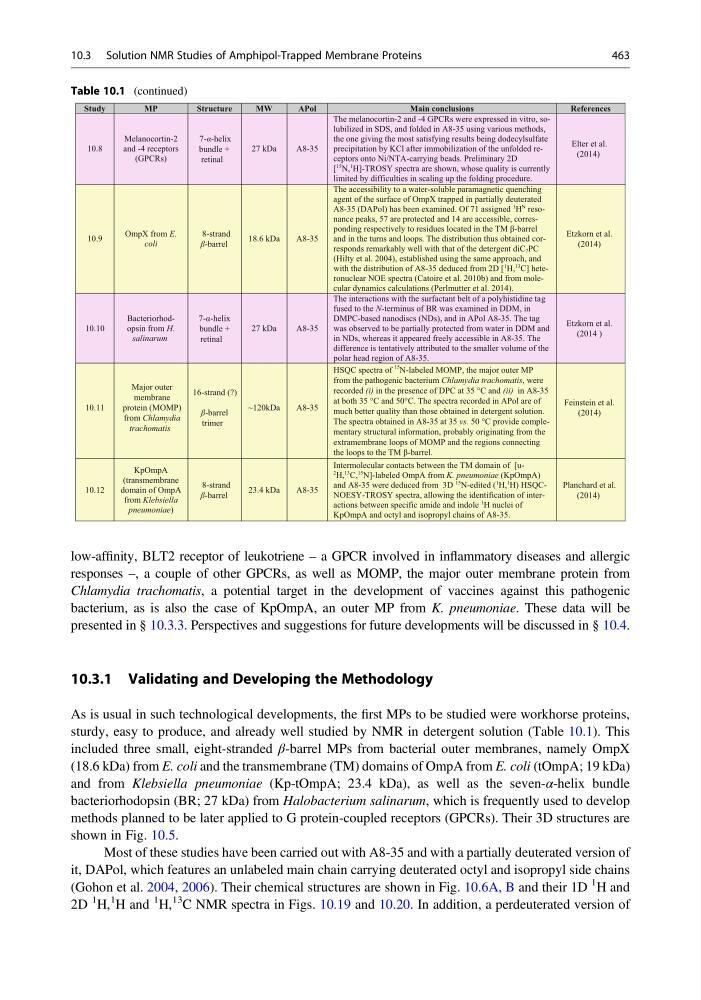
sturdy, easy to produce, and already well studied by NMR in detergent solution (Table 10.1). This
included three small, eight-stranded β-barrel MPs from bacterial outer membranes, namely OmpX
(18.6 kDa) from E. coli and the transmembrane (TM) domains of OmpA from E. coli (tOmpA; 19 kDa)
and from Klebsiella pneumoniae (Kp-tOmpA; 23.4 kDa), as well as the seven-α-helix bundle
bacteriorhodopsin (BR; 27 kDa) from Halobacterium salinarum, which is frequently used to develop
methods planned to be later applied to G protein-coupled receptors (GPCRs). Their 3D structures are
shown in Fig. 10.5.
Most of these studies have been carried out with A8-35 and with a partially deuterated version of
it, DAPol, which features an unlabeled main chain carrying deuterated octyl and isopropyl side chains
(Gohon et al. 2004, 2006). Their chemical structures are shown in Fig. 10.6A, B and their 1D
1 H and
2D
1 H,
1 H and
1 H,
13 C NMR spectra in Figs. 10.19 and 10.20. In addition, a perdeuterated version of
Table 10.1 (continued)
10.8
Melanocortin-2
and -4 receptors
(GPCRs)
7-α-helix
bundle +
retinal
27 kDa
A8-35
The melanocortin-2 and -4 GPCRs were expressed in vitro, solubilized in SDS, and folded in A8-35 using various methods,
the one giving the most satisfying results being dodecylsulfate
precipitation by KCl after immobilization of the unfolded receptors onto Ni/NTA-carrying beads. Preliminary 2D
[
15 N,
1 H]-TROSY spectra are shown, whose quality is currently
limited by difficulties in scaling up the folding procedure.
Elter et al.
(2014)
10.9
OmpX from E.
coli
8-strand
β-barrel
18.6 kDa
A8-35
The accessibility to a water-soluble paramagnetic quenching
agent of the surface of OmpX trapped in partially deuterated
A8-35 (DAPol) has been examined. Of 71 assigned
1 H
N resonance peaks, 57 are protected and 14 are accessible, corresponding respectively to residues located in the TM β-barrel
and in the turns and loops. The distribution thus obtained corresponds remarkably well with that of the detergent diC7PC
(Hilty et al. 2004), established using the same approach, and
with the distribution of A8-35 deduced from 2D [
1 H,
13 C] heteronuclear NOE spectra (Catoire et al. 2010b) and from molecular dynamics calculations (Perlmutter et al. 2014).
Etzkorn et al.
(2014)
10.10
Bacteriorhodopsin from H.
salinarum
7-α-helix
bundle +
retinal
27 kDa
A8-35
The interactions with the surfactant belt of a polyhistidine tag
fused to the N-terminus of BR was examined in DDM, in
DMPC-based nanodiscs (NDs), and in APol A8-35. The tag
was observed to be partially protected from water in DDM and
in NDs, whereas it appeared freely accessible in A8-35. The
difference is tentatively attributed to the smaller volume of the
polar head region of A8-35.
Etzkorn et al.
(2014 )
10.11
Major outer
membrane
protein (MOMP)
from Chlamydia
trachomatis
~120kDa
A8-35
HSQC spectra of
15
N-labeled MOMP, the major outer MP
from the pathogenic bacterium Chlamydia trachomatis, were
recorded (i) in the presence of DPC at 35 °C and (ii) in A8-35
at both 35 °C and 50°C. The spectra recorded in APol are of
much better quality than those obtained in detergent solution.
The spectra obtained in A8-35 at 35 vs. 50 °C provide complementary structural information, probably originating from the
extramembrane loops of MOMP and the regions connecting
the loops to the TM β-barrel.
Feinstein et al.
(2014)
16-strand (?)
β-barrel
trimer
10.12
KpOmpA
(transmembrane
domain of OmpA
from Klebsiella
pneumoniae)
8-strand
β-barrel
23.4 kDa
A8-35
Intermolecular contacts between the TM domain of [u2 H,
13 C,
15 N]-labeled OmpA from K. pneumoniae (KpOmpA)
and A8-35 were deduced from 3D
15 N-edited (
1 H,
1 H) HSQCNOESY-TROSY spectra, allowing the identification of interactions between specific amide and indole
1 H nuclei of
KpOmpA and octyl and isopropyl chains of A8-35.
Planchard et al.
(2014)
Study
MP
Structure
MW
APol
Main conclusions
References
10.3 Solution NMR Studies of Amphipol-Trapped Membrane Proteins
463
responses –, a couple of other GPCRs, as well as MOMP, the major outer membrane protein from
Chlamydia trachomatis, a potential target in the development of vaccines against this pathogenic
bacterium, as is also the case of KpOmpA, an outer MP from K. pneumoniae. These data will be
presented in § 10.3.3. Perspectives and suggestions for future developments will be discussed in § 10.4.
10.3.1 Validating and Developing the Methodology
As is usual in such technological developments, the first MPs to be studied were workhorse proteins,
sturdy, easy to produce, and already well studied by NMR in detergent solution (Table 10.1). This
included three small, eight-stranded β-barrel MPs from bacterial outer membranes, namely OmpX
(18.6 kDa) from E. coli and the transmembrane (TM) domains of OmpA from E. coli (tOmpA; 19 kDa)
and from Klebsiella pneumoniae (Kp-tOmpA; 23.4 kDa), as well as the seven-α-helix bundle
bacteriorhodopsin (BR; 27 kDa) from Halobacterium salinarum, which is frequently used to develop
methods planned to be later applied to G protein-coupled receptors (GPCRs). Their 3D structures are
shown in Fig. 10.5.
Most of these studies have been carried out with A8-35 and with a partially deuterated version of
it, DAPol, which features an unlabeled main chain carrying deuterated octyl and isopropyl side chains
(Gohon et al. 2004, 2006). Their chemical structures are shown in Fig. 10.6A, B and their 1D
1 H and
2D
1 H,
1 H and
1 H,
13 C NMR spectra in Figs. 10.19 and 10.20. In addition, a perdeuterated version of
Table 10.1 (continued)
10.8
Melanocortin-2
and -4 receptors
(GPCRs)
7-α-helix
bundle +
retinal
27 kDa
A8-35
The melanocortin-2 and -4 GPCRs were expressed in vitro, solubilized in SDS, and folded in A8-35 using various methods,
the one giving the most satisfying results being dodecylsulfate
precipitation by KCl after immobilization of the unfolded receptors onto Ni/NTA-carrying beads. Preliminary 2D
[
15 N,
1 H]-TROSY spectra are shown, whose quality is currently
limited by difficulties in scaling up the folding procedure.
Elter et al.
(2014)
10.9
OmpX from E.
coli
8-strand
β-barrel
18.6 kDa
A8-35
The accessibility to a water-soluble paramagnetic quenching
agent of the surface of OmpX trapped in partially deuterated
A8-35 (DAPol) has been examined. Of 71 assigned
1 H
N resonance peaks, 57 are protected and 14 are accessible, corresponding respectively to residues located in the TM β-barrel
and in the turns and loops. The distribution thus obtained corresponds remarkably well with that of the detergent diC7PC
(Hilty et al. 2004), established using the same approach, and
with the distribution of A8-35 deduced from 2D [
1 H,
13 C] heteronuclear NOE spectra (Catoire et al. 2010b) and from molecular dynamics calculations (Perlmutter et al. 2014).
Etzkorn et al.
(2014)
10.10
Bacteriorhodopsin from H.
salinarum
7-α-helix
bundle +
retinal
27 kDa
A8-35
The interactions with the surfactant belt of a polyhistidine tag
fused to the N-terminus of BR was examined in DDM, in
DMPC-based nanodiscs (NDs), and in APol A8-35. The tag
was observed to be partially protected from water in DDM and
in NDs, whereas it appeared freely accessible in A8-35. The
difference is tentatively attributed to the smaller volume of the
polar head region of A8-35.
Etzkorn et al.
(2014 )
10.11
Major outer
membrane
protein (MOMP)
from Chlamydia
trachomatis
~120kDa
A8-35
HSQC spectra of
15
N-labeled MOMP, the major outer MP
from the pathogenic bacterium Chlamydia trachomatis, were
recorded (i) in the presence of DPC at 35 °C and (ii) in A8-35
at both 35 °C and 50°C. The spectra recorded in APol are of
much better quality than those obtained in detergent solution.
The spectra obtained in A8-35 at 35 vs. 50 °C provide complementary structural information, probably originating from the
extramembrane loops of MOMP and the regions connecting
the loops to the TM β-barrel.
Feinstein et al.
(2014)
16-strand (?)
β-barrel
trimer
10.12
KpOmpA
(transmembrane
domain of OmpA
from Klebsiella
pneumoniae)
8-strand
β-barrel
23.4 kDa
A8-35
Intermolecular contacts between the TM domain of [u2 H,
13 C,
15 N]-labeled OmpA from K. pneumoniae (KpOmpA)
and A8-35 were deduced from 3D
15 N-edited (
1 H,
1 H) HSQCNOESY-TROSY spectra, allowing the identification of interactions between specific amide and indole
1 H nuclei of
KpOmpA and octyl and isopropyl chains of A8-35.
Planchard et al.
(2014)
Study
MP
Structure
MW
APol
Main conclusions
References
10.3 Solution NMR Studies of Amphipol-Trapped Membrane Proteins
463