

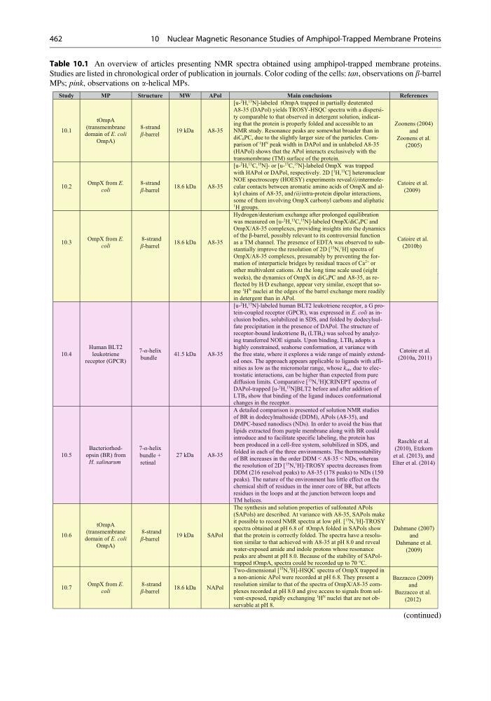
Table 10.1 An overview of articles presenting NMR spectra obtained using amphipol-trapped membrane proteins.
Studies are listed in chronological order of publication in journals. Color coding of the cells: tan, observations on b-barrel
MPs; pink, observations on a-helical MPs.
weeks), the dynamics of OmpX in diC6PC and A8-35, as reflected by H/D exchange, appear very similar, except that some
1 H
N nuclei at the edges of the barrel exchange more readily
in detergent than in APol.
10.4
Human BLT2
leukotriene
receptor (GPCR)
7-α-helix
bundle
41.5 kDa
A8-35
[u2 H,
15 N]-labeled human BLT2 leukotriene receptor, a G protein-coupled receptor (GPCR), was expressed in E. coli as inclusion bodies, solubilized in SDS, and folded by dodecylsulfate precipitation in the presence of DAPol. The structure of
receptor-bound leukotriene B4 (LTB4) was solved by analyzing transferred NOE signals. Upon binding, LTB4 adopts a
highly constrained, seahorse conformation, at variance with
the free state, where it explores a wide range of mainly extended ones. The approach appears applicable to ligands with affinities as low as the micromolar range, whose kon, due to electrostatic interactions, can be higher than expected from pure
diffusion limits. Comparative [
15 N,
1 H]CRINEPT spectra of
DAPol-trapped [u2 H,
15 N]BLT2 before and after addition of
LTB4 show that binding of the ligand induces conformational
changes in the receptor.
Catoire et al.
(2010a, 2011)
10.5
Bacteriorhodopsin (BR) from
H. salinarum
7-α-helix
bundle +
retinal
27 kDa
A8-35
A detailed comparison is presented of solution NMR studies
of BR in dodecylmaltoside (DDM), APols (A8-35), and
DMPC-based nanodiscs (NDs). In order to avoid the bias that
lipids extracted from purple membrane along with BR could
introduce and to facilitate specific labeling, the protein has
been produced in a cell-free system, solubilized in SDS, and
folded in each of the three environments. The thermostability
of BR increases in the order DDM < A8-35 < NDs, whereas
the resolution of 2D [
15 N,
1 H]-TROSY spectra decreases from
DDM (216 resolved peaks) to A8-35 (178 peaks) to NDs (150
peaks). The nature of the environment has little effect on the
chemical shift of residues in the inner core of BR, but affects
residues in the loops and at the junction between loops and
TM helices.
Raschle et al.
(2010), Etzkorn
et al. (2013), and
Elter et al. (2014)
10.6
tOmpA
(transmembrane
domain of E. coli
OmpA)
8-strand
β-barrel
19 kDa
SAPol
The synthesis and solution properties of sulfonated APols
(SAPols) are described. At variance with A8-35, SAPols make
it possible to record NMR spectra at low pH. [
15 N,
1 H]-TROSY
spectra obtained at pH 6.8 of tOmpA folded in SAPols show
that the protein is correctly folded. The spectra have a resolution similar to that achieved with A8-35 at pH 8.0 and reveal
water-exposed amide and indole protons whose resonance
peaks are absent at pH 8.0. Because of the stability of SAPoltrapped tOmpA, spectra could be recorded up to 70 °C.
Dahmane (2007)
and
Dahmane et al.
(2009)
10.7
OmpX from E.
coli
8-strand
β-barrel
18.6 kDa
NAPol
Two-dimensional [
15 N,
1 H]-HSQC spectra of OmpX trapped in
a non-anionic APol were recorded at pH 6.8. They present a
resolution similar to that of the spectra of OmpX/A8-35 complexes recorded at pH 8.0 and give access to signals from solvent-exposed, rapidly exchanging
1 H
N nuclei that are not observable at pH 8.
Bazzacco (2009)
and
Bazzacco et al.
(2012)
Study
MP
Structure
MW
APol
Main conclusions
References
10.1
tOmpA
(transmembrane
domain of E. coli
OmpA)
8-strand
β-barrel
19 kDa
A8-35
[u2 H,
15 N]-labeled tOmpA trapped in partially deuterated
A8-35 (DAPol) yields TROSY-HSQC spectra with a dispersity comparable to that observed in detergent solution, indicating that the protein is properly folded and accessible to an
NMR study. Resonance peaks are somewhat broader than in
diC6PC, due to the slightly larger size of the particles. Comparison of
1 H
N peak width in DAPol and in unlabeled A8-35
(HAPol) shows that the APol interacts exclusively with the
transmembrane (TM) surface of the protein.
Zoonens (2004)
and
Zoonens et al.
(2005)
10.2
OmpX from E.
coli
8-strand
β-barrel
18.6 kDa
A8-35
[u2 H,
13 C,
15 N]- or [u13 C,
15 N]-labeled OmpX was trapped
with HAPol or DAPol, respectively. 2D [
1 H,
13 C] heteronuclear
NOE spectroscopy (HOESY) experiments reveal (i) intermolecular contacts between aromatic amino acids of OmpX and alkyl chains of A8-35, and (ii) intra-protein dipolar interactions,
some of them involving OmpX carbonyl carbons and aliphatic
1 H groups.
Catoire et al.
(2009)
10.3
OmpX from E.
coli
8-strand
β-barrel
18.6 kDa
A8-35
Hydrogen/deuterium exchange after prolonged equilibration
was measured on [u2 H,
13 C,
15 N]-labeled OmpX/diC6PC and
OmpX/A8-35 complexes, providing insights into the dynamics
of the β-barrel, possibly relevant to its controversial function
as a TM channel. The presence of EDTA was observed to substantially improve the resolution of 2D [
15 N,
1 H] spectra of
OmpX/A8-35 complexes, presumably by preventing the formation of interparticle bridges by residual traces of Ca
2+ or
other multivalent cations. At the long time scale used (eight
Catoire et al.
(2010b)
(continued)
462
10 Nuclear Magnetic Resonance Studies of Amphipol-Trapped Membrane Proteins
Studies are listed in chronological order of publication in journals. Color coding of the cells: tan, observations on b-barrel
MPs; pink, observations on a-helical MPs.
weeks), the dynamics of OmpX in diC6PC and A8-35, as reflected by H/D exchange, appear very similar, except that some
1 H
N nuclei at the edges of the barrel exchange more readily
in detergent than in APol.
10.4
Human BLT2
leukotriene
receptor (GPCR)
7-α-helix
bundle
41.5 kDa
A8-35
[u2 H,
15 N]-labeled human BLT2 leukotriene receptor, a G protein-coupled receptor (GPCR), was expressed in E. coli as inclusion bodies, solubilized in SDS, and folded by dodecylsulfate precipitation in the presence of DAPol. The structure of
receptor-bound leukotriene B4 (LTB4) was solved by analyzing transferred NOE signals. Upon binding, LTB4 adopts a
highly constrained, seahorse conformation, at variance with
the free state, where it explores a wide range of mainly extended ones. The approach appears applicable to ligands with affinities as low as the micromolar range, whose kon, due to electrostatic interactions, can be higher than expected from pure
diffusion limits. Comparative [
15 N,
1 H]CRINEPT spectra of
DAPol-trapped [u2 H,
15 N]BLT2 before and after addition of
LTB4 show that binding of the ligand induces conformational
changes in the receptor.
Catoire et al.
(2010a, 2011)
10.5
Bacteriorhodopsin (BR) from
H. salinarum
7-α-helix
bundle +
retinal
27 kDa
A8-35
A detailed comparison is presented of solution NMR studies
of BR in dodecylmaltoside (DDM), APols (A8-35), and
DMPC-based nanodiscs (NDs). In order to avoid the bias that
lipids extracted from purple membrane along with BR could
introduce and to facilitate specific labeling, the protein has
been produced in a cell-free system, solubilized in SDS, and
folded in each of the three environments. The thermostability
of BR increases in the order DDM < A8-35 < NDs, whereas
the resolution of 2D [
15 N,
1 H]-TROSY spectra decreases from
DDM (216 resolved peaks) to A8-35 (178 peaks) to NDs (150
peaks). The nature of the environment has little effect on the
chemical shift of residues in the inner core of BR, but affects
residues in the loops and at the junction between loops and
TM helices.
Raschle et al.
(2010), Etzkorn
et al. (2013), and
Elter et al. (2014)
10.6
tOmpA
(transmembrane
domain of E. coli
OmpA)
8-strand
β-barrel
19 kDa
SAPol
The synthesis and solution properties of sulfonated APols
(SAPols) are described. At variance with A8-35, SAPols make
it possible to record NMR spectra at low pH. [
15 N,
1 H]-TROSY
spectra obtained at pH 6.8 of tOmpA folded in SAPols show
that the protein is correctly folded. The spectra have a resolution similar to that achieved with A8-35 at pH 8.0 and reveal
water-exposed amide and indole protons whose resonance
peaks are absent at pH 8.0. Because of the stability of SAPoltrapped tOmpA, spectra could be recorded up to 70 °C.
Dahmane (2007)
and
Dahmane et al.
(2009)
10.7
OmpX from E.
coli
8-strand
β-barrel
18.6 kDa
NAPol
Two-dimensional [
15 N,
1 H]-HSQC spectra of OmpX trapped in
a non-anionic APol were recorded at pH 6.8. They present a
resolution similar to that of the spectra of OmpX/A8-35 complexes recorded at pH 8.0 and give access to signals from solvent-exposed, rapidly exchanging
1 H
N nuclei that are not observable at pH 8.
Bazzacco (2009)
and
Bazzacco et al.
(2012)
Study
MP
Structure
MW
APol
Main conclusions
References
10.1
tOmpA
(transmembrane
domain of E. coli
OmpA)
8-strand
β-barrel
19 kDa
A8-35
[u2 H,
15 N]-labeled tOmpA trapped in partially deuterated
A8-35 (DAPol) yields TROSY-HSQC spectra with a dispersity comparable to that observed in detergent solution, indicating that the protein is properly folded and accessible to an
NMR study. Resonance peaks are somewhat broader than in
diC6PC, due to the slightly larger size of the particles. Comparison of
1 H
N peak width in DAPol and in unlabeled A8-35
(HAPol) shows that the APol interacts exclusively with the
transmembrane (TM) surface of the protein.
Zoonens (2004)
and
Zoonens et al.
(2005)
10.2
OmpX from E.
coli
8-strand
β-barrel
18.6 kDa
A8-35
[u2 H,
13 C,
15 N]- or [u13 C,
15 N]-labeled OmpX was trapped
with HAPol or DAPol, respectively. 2D [
1 H,
13 C] heteronuclear
NOE spectroscopy (HOESY) experiments reveal (i) intermolecular contacts between aromatic amino acids of OmpX and alkyl chains of A8-35, and (ii) intra-protein dipolar interactions,
some of them involving OmpX carbonyl carbons and aliphatic
1 H groups.
Catoire et al.
(2009)
10.3
OmpX from E.
coli
8-strand
β-barrel
18.6 kDa
A8-35
Hydrogen/deuterium exchange after prolonged equilibration
was measured on [u2 H,
13 C,
15 N]-labeled OmpX/diC6PC and
OmpX/A8-35 complexes, providing insights into the dynamics
of the β-barrel, possibly relevant to its controversial function
as a TM channel. The presence of EDTA was observed to substantially improve the resolution of 2D [
15 N,
1 H] spectra of
OmpX/A8-35 complexes, presumably by preventing the formation of interparticle bridges by residual traces of Ca
2+ or
other multivalent cations. At the long time scale used (eight
Catoire et al.
(2010b)
(continued)
462
10 Nuclear Magnetic Resonance Studies of Amphipol-Trapped Membrane Proteins