

sedimentation coefficients depend on the ionic strength. Cryo-EM single-particle imaging revealed that
the doublet is formed by two AcrB/SMALP complexes associated via their SMALP belts (see
Chap. 12, Fig. 12.15).
9.3.8
Small-Angle X-ray and Neutron Solution Scattering
9.3.8.1 Introduction
Radiation scattering is a powerful approach to examining the mass, size, shape, and internal organization of solutes, as well as interactions between them. In addition to five DLS studies, which will be
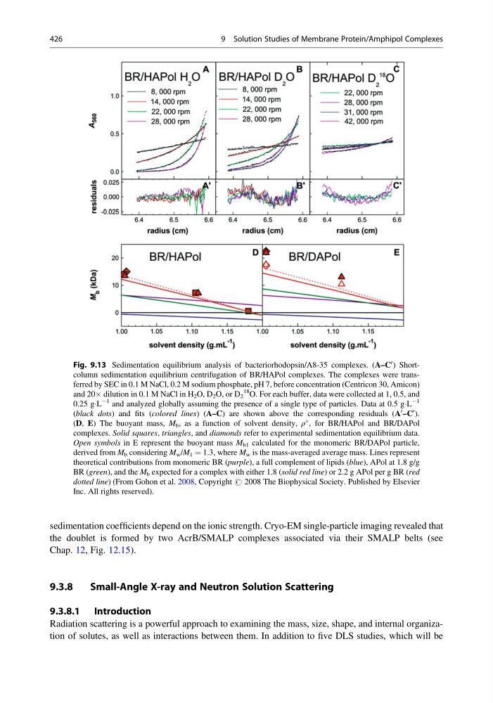
Fig. 9.13 Sedimentation equilibrium analysis of bacteriorhodopsin/A8-35 complexes. (A–C
0 ) Shortcolumn sedimentation equilibrium centrifugation of BR/HAPol complexes. The complexes were transferred by SEC in 0.1 M NaCl, 0.2 M sodium phosphate, pH 7, before concentration (Centricon 30, Amicon)
and 20Â dilution in 0.1 M NaCl in H 2 O, D 2 O, or D 2
18
O. For each buffer, data were collected at 1, 0.5, and
0.25 gÁL
À1 and analyzed globally assuming the presence of a single type of particles. Data at 0.5 gÁL
À1
(black dots) and fits (colored lines) (A–C) are shown above the corresponding residuals (A
0 –C
0 ).
(D, E) The buoyant mass, M b , as a function of solvent density, ρ
, for BR/HAPol and BR/DAPol
complexes. Solid squares, triangles, and diamonds refer to experimental sedimentation equilibrium data.
Open symbols in E represent the buoyant mass M b1 calculated for the monomeric BR/DAPol particle,
derived from M b considering M w /M 1 ¼ 1.3, where M w is the mass-averaged average mass. Lines represent
theoretical contributions from monomeric BR (purple), a full complement of lipids (blue), APol at 1.8 g/g
BR (green), and the M b expected for a complex with either 1.8 (solid red line) or 2.2 g APol per g BR (red
dotted line) (From Gohon et al. 2008, Copyright # 2008 The Biophysical Society. Published by Elsevier
Inc. All rights reserved).
426
9 Solution Studies of Membrane Protein/Amphipol Complexes
the doublet is formed by two AcrB/SMALP complexes associated via their SMALP belts (see
Chap. 12, Fig. 12.15).
9.3.8
Small-Angle X-ray and Neutron Solution Scattering
9.3.8.1 Introduction
Radiation scattering is a powerful approach to examining the mass, size, shape, and internal organization of solutes, as well as interactions between them. In addition to five DLS studies, which will be
Fig. 9.13 Sedimentation equilibrium analysis of bacteriorhodopsin/A8-35 complexes. (A–C
0 ) Shortcolumn sedimentation equilibrium centrifugation of BR/HAPol complexes. The complexes were transferred by SEC in 0.1 M NaCl, 0.2 M sodium phosphate, pH 7, before concentration (Centricon 30, Amicon)
and 20Â dilution in 0.1 M NaCl in H 2 O, D 2 O, or D 2
18
O. For each buffer, data were collected at 1, 0.5, and
0.25 gÁL
À1 and analyzed globally assuming the presence of a single type of particles. Data at 0.5 gÁL
À1
(black dots) and fits (colored lines) (A–C) are shown above the corresponding residuals (A
0 –C
0 ).
(D, E) The buoyant mass, M b , as a function of solvent density, ρ
, for BR/HAPol and BR/DAPol
complexes. Solid squares, triangles, and diamonds refer to experimental sedimentation equilibrium data.
Open symbols in E represent the buoyant mass M b1 calculated for the monomeric BR/DAPol particle,
derived from M b considering M w /M 1 ¼ 1.3, where M w is the mass-averaged average mass. Lines represent
theoretical contributions from monomeric BR (purple), a full complement of lipids (blue), APol at 1.8 g/g
BR (green), and the M b expected for a complex with either 1.8 (solid red line) or 2.2 g APol per g BR (red
dotted line) (From Gohon et al. 2008, Copyright # 2008 The Biophysical Society. Published by Elsevier
Inc. All rights reserved).
426
9 Solution Studies of Membrane Protein/Amphipol Complexes