

water molecules that hydrate alkaline cations in the bulk and, by their geometry, define the specificity
of the channel (Fig. 1.10C).
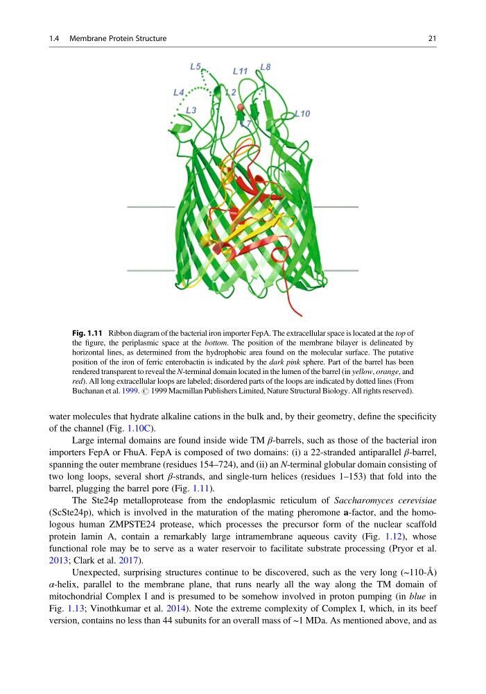
Large internal domains are found inside wide TM β-barrels, such as those of the bacterial iron
importers FepA or FhuA. FepA is composed of two domains: (i) a 22-stranded antiparallel β-barrel,
spanning the outer membrane (residues 154–724), and (ii) an N-terminal globular domain consisting of
two long loops, several short β-strands, and single-turn helices (residues 1–153) that fold into the
barrel, plugging the barrel pore (Fig. 1.11).
The Ste24p metalloprotease from the endoplasmic reticulum of Saccharomyces cerevisiae
(ScSte24p), which is involved in the maturation of the mating pheromone a-factor, and the homologous human ZMPSTE24 protease, which processes the precursor form of the nuclear scaffold
protein lamin A, contain a remarkably large intramembrane aqueous cavity (Fig. 1.12), whose
functional role may be to serve as a water reservoir to facilitate substrate processing (Pryor et al.
2013; Clark et al. 2017).
Unexpected, surprising structures continue to be discovered, such as the very long (~110-Å)
α-helix, parallel to the membrane plane, that runs nearly all the way along the TM domain of
mitochondrial Complex I and is presumed to be somehow involved in proton pumping (in blue in
Fig. 1.13; Vinothkumar et al. 2014). Note the extreme complexity of Complex I, which, in its beef
version, contains no less than 44 subunits for an overall mass of ~1 MDa. As mentioned above, and as
Fig. 1.11 Ribbon diagram of the bacterial iron importer FepA. The extracellular space is located at the top of
the figure, the periplasmic space at the bottom. The position of the membrane bilayer is delineated by
horizontal lines, as determined from the hydrophobic area found on the molecular surface. The putative
position of the iron of ferric enterobactin is indicated by the dark pink sphere. Part of the barrel has been
rendered transparent to reveal the N-terminal domain located in the lumen of the barrel (in yellow, orange, and
red). All long extracellular loops are labeled; disordered parts of the loops are indicated by dotted lines (From
Buchanan et al. 1999. # 1999 Macmillan Publishers Limited, Nature Structural Biology. All rights reserved).
1.4 Membrane Protein Structure
21
of the channel (Fig. 1.10C).
Large internal domains are found inside wide TM β-barrels, such as those of the bacterial iron
importers FepA or FhuA. FepA is composed of two domains: (i) a 22-stranded antiparallel β-barrel,
spanning the outer membrane (residues 154–724), and (ii) an N-terminal globular domain consisting of
two long loops, several short β-strands, and single-turn helices (residues 1–153) that fold into the
barrel, plugging the barrel pore (Fig. 1.11).
The Ste24p metalloprotease from the endoplasmic reticulum of Saccharomyces cerevisiae
(ScSte24p), which is involved in the maturation of the mating pheromone a-factor, and the homologous human ZMPSTE24 protease, which processes the precursor form of the nuclear scaffold
protein lamin A, contain a remarkably large intramembrane aqueous cavity (Fig. 1.12), whose
functional role may be to serve as a water reservoir to facilitate substrate processing (Pryor et al.
2013; Clark et al. 2017).
Unexpected, surprising structures continue to be discovered, such as the very long (~110-Å)
α-helix, parallel to the membrane plane, that runs nearly all the way along the TM domain of
mitochondrial Complex I and is presumed to be somehow involved in proton pumping (in blue in
Fig. 1.13; Vinothkumar et al. 2014). Note the extreme complexity of Complex I, which, in its beef
version, contains no less than 44 subunits for an overall mass of ~1 MDa. As mentioned above, and as
Fig. 1.11 Ribbon diagram of the bacterial iron importer FepA. The extracellular space is located at the top of
the figure, the periplasmic space at the bottom. The position of the membrane bilayer is delineated by
horizontal lines, as determined from the hydrophobic area found on the molecular surface. The putative
position of the iron of ferric enterobactin is indicated by the dark pink sphere. Part of the barrel has been
rendered transparent to reveal the N-terminal domain located in the lumen of the barrel (in yellow, orange, and
red). All long extracellular loops are labeled; disordered parts of the loops are indicated by dotted lines (From
Buchanan et al. 1999. # 1999 Macmillan Publishers Limited, Nature Structural Biology. All rights reserved).
1.4 Membrane Protein Structure
21