

will be described in more detail in the chapter dedicated to electron microscopy (Chap. 12, § 12.3.3),
Complex I is itself integrated into an even larger ensemble, the respirasome (mitochondrial
supercomplex B), where it is associated to a dimer of Complex III (the cytochrome bc 1 complex)
and a monomer of Complex IV (the cytochrome c oxidase) (see Fig. 12.17). This can serve as a
reminder that, when we handle “a membrane protein” in solution, as will be described in the next
chapter, it may well be only a fraction of a larger ensemble that has not resisted solubilization (see
e.g. Kühlbrandt 2015).
1.5
Membrane Protein/Lipid Interactions
1.5.1
The Fluid Mosaic Model
At the end of the 1940s, biological membranes were known to comprise both lipids and proteins, and
the ability of lipids to spontaneously organize into bilayers had long been recognized. Nothing
however was known of the sequence nor the 3D structure of MPs nor of their arrangement respective
to lipids. A long controversy ensued (see e.g. Green et al. 1967), which was resolved, in the early
1970s, in favor of the fluid mosaic model proposed by Seymour J. Singer and Garth L. Nicolson
(Singer and Nicolson 1972). According to this model, “the bulk of the phospholipid is organized as a
discontinuous, fluid bilayer, although a small fraction of the lipid may interact specifically with the
membrane proteins. The fluid mosaic structure is therefore formally analogous to a two-dimensional
oriented solution of integral proteins (or lipoproteins) in the viscous phospholipid bilayer solvent”
(Fig. 1.14A). Important in the elaboration of this model were the realization that a continuous (but for
the inserted proteins) bilayer of lipids would provide the hydrophobic barrier needed to maintain
compositional differences between the aqueous compartments separated by membranes and the fact
that membrane proteins (or, at least, some of them) could redistribute rapidly in the membrane plane
when, for instance, they were cross-linked by antibodies.
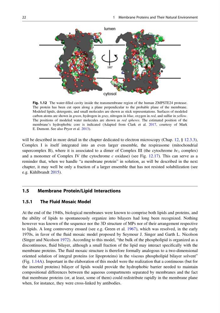
Fig. 1.12 The water-filled cavity inside the transmembrane region of the human ZMPSTE24 protease.
The protein has been cut open along a plane perpendicular to the probable plane of the membrane.
Modeled lipids, detergents, and small molecules are shown as stick representations. Surfaces of modeled
carbon atoms are shown in green, hydrogen in gray, nitrogen in blue, oxygen in red, and sulfur in yellow.
The positions of modeled water molecules are shown as red spheres. The estimated position of the
membrane’s hydrophobic core is indicated (Adapted from Clark et al. 2017, courtesy of Mark
E. Dumont. See also Pryor et al. 2013).
22
1 Membrane Proteins and Their Natural Environment
Complex I is itself integrated into an even larger ensemble, the respirasome (mitochondrial
supercomplex B), where it is associated to a dimer of Complex III (the cytochrome bc 1 complex)
and a monomer of Complex IV (the cytochrome c oxidase) (see Fig. 12.17). This can serve as a
reminder that, when we handle “a membrane protein” in solution, as will be described in the next
chapter, it may well be only a fraction of a larger ensemble that has not resisted solubilization (see
e.g. Kühlbrandt 2015).
1.5
Membrane Protein/Lipid Interactions
1.5.1
The Fluid Mosaic Model
At the end of the 1940s, biological membranes were known to comprise both lipids and proteins, and
the ability of lipids to spontaneously organize into bilayers had long been recognized. Nothing
however was known of the sequence nor the 3D structure of MPs nor of their arrangement respective
to lipids. A long controversy ensued (see e.g. Green et al. 1967), which was resolved, in the early
1970s, in favor of the fluid mosaic model proposed by Seymour J. Singer and Garth L. Nicolson
(Singer and Nicolson 1972). According to this model, “the bulk of the phospholipid is organized as a
discontinuous, fluid bilayer, although a small fraction of the lipid may interact specifically with the
membrane proteins. The fluid mosaic structure is therefore formally analogous to a two-dimensional
oriented solution of integral proteins (or lipoproteins) in the viscous phospholipid bilayer solvent”
(Fig. 1.14A). Important in the elaboration of this model were the realization that a continuous (but for
the inserted proteins) bilayer of lipids would provide the hydrophobic barrier needed to maintain
compositional differences between the aqueous compartments separated by membranes and the fact
that membrane proteins (or, at least, some of them) could redistribute rapidly in the membrane plane
when, for instance, they were cross-linked by antibodies.
Fig. 1.12 The water-filled cavity inside the transmembrane region of the human ZMPSTE24 protease.
The protein has been cut open along a plane perpendicular to the probable plane of the membrane.
Modeled lipids, detergents, and small molecules are shown as stick representations. Surfaces of modeled
carbon atoms are shown in green, hydrogen in gray, nitrogen in blue, oxygen in red, and sulfur in yellow.
The positions of modeled water molecules are shown as red spheres. The estimated position of the
membrane’s hydrophobic core is indicated (Adapted from Clark et al. 2017, courtesy of Mark
E. Dumont. See also Pryor et al. 2013).
22
1 Membrane Proteins and Their Natural Environment