

Similarly, in ion channels such as the potassium channel KcsA from Streptomyces lividans
(Doyle et al. 1998), the continuous fence of TM helices formed by the assembly of four two-TM helix
monomers protects a central TM aqueous channel (Fig. 1.10). The channel contains a reentrant loop in
which four extended strands provide the main chain oxygen atoms that can substitute for some of the
Fig. 1.9 Transmembrane arrangement of a bacterial outer membrane porin, OmpF from E. coli. (A)
General fold of a porin monomer. The large hollow β-barrel structure is formed by the antiparallel
arrangement of 16 β-strands. The strands are connected by short loops or regular turns on the periplasmic
rim (bottom), whereas long irregular loops face the cell exterior (top). The internal loop, which connects
β-strands 5 and 6 and plunges inside the barrel, is highlighted in dark. The chain N- and C-termini interact
one with another. The surface closest to the viewer, which faces the threefold symmetry axis of the trimer,
is involved in subunit-subunit contacts. The approximate position of the membrane hydrophobic core is
indicated (X-ray structure from Cowan et al. 1992). (B) Schematic representation of the OmpF trimer. The
view is from the extracellular space along the molecular threefold symmetry axis. (C) Slice through the
center of the OmpF trimer structure, represented by a stick model. The molecular surface is indicated so as
to visualize more clearly the three pores (about 7 Â 11 Å in size). Same view as in B. (Adapted from
Schirmer 1998. # 1998 Academic Press. All rights reserved).
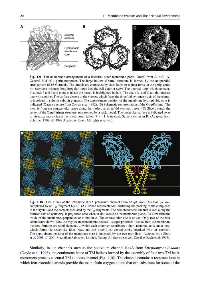
Fig. 1.10 Two views of the tetrameric KcsA potassium channel from Streptomyces lividans (yellow)
complexed by an F ab fragment (cyan). (A) Ribbon representation illustrating the packing of the complexes
in the crystals and the contacts mediated by the F ab fragments. The homotetrameric channel is seen along the
fourfold axis of symmetry, in projection onto what, in situ, would be the membrane plane. (B) View from the
inside of the membrane, perpendicular to that in A. The extracellular side is on top. Only two of the four
subunits are shown. Note the way the transmembrane helices – two per protomer – isolate from the membrane
the pore-forming structural elements, to which each protomer contributes a short, reentrant helix and a loop,
which forms the selectivity filter (red), and the water-filled central cavity (marked with an asterisk).
The approximate position of the membrane core is indicated by the two gray lines (Adapted from Zhou
et al. 2001. # 2001 Macmillan Publishers Limited, Nature. All rights reserved. See also Doyle et al. 1998).
20
1 Membrane Proteins and Their Natural Environment
(Doyle et al. 1998), the continuous fence of TM helices formed by the assembly of four two-TM helix
monomers protects a central TM aqueous channel (Fig. 1.10). The channel contains a reentrant loop in
which four extended strands provide the main chain oxygen atoms that can substitute for some of the
Fig. 1.9 Transmembrane arrangement of a bacterial outer membrane porin, OmpF from E. coli. (A)
General fold of a porin monomer. The large hollow β-barrel structure is formed by the antiparallel
arrangement of 16 β-strands. The strands are connected by short loops or regular turns on the periplasmic
rim (bottom), whereas long irregular loops face the cell exterior (top). The internal loop, which connects
β-strands 5 and 6 and plunges inside the barrel, is highlighted in dark. The chain N- and C-termini interact
one with another. The surface closest to the viewer, which faces the threefold symmetry axis of the trimer,
is involved in subunit-subunit contacts. The approximate position of the membrane hydrophobic core is
indicated (X-ray structure from Cowan et al. 1992). (B) Schematic representation of the OmpF trimer. The
view is from the extracellular space along the molecular threefold symmetry axis. (C) Slice through the
center of the OmpF trimer structure, represented by a stick model. The molecular surface is indicated so as
to visualize more clearly the three pores (about 7 Â 11 Å in size). Same view as in B. (Adapted from
Schirmer 1998. # 1998 Academic Press. All rights reserved).
Fig. 1.10 Two views of the tetrameric KcsA potassium channel from Streptomyces lividans (yellow)
complexed by an F ab fragment (cyan). (A) Ribbon representation illustrating the packing of the complexes
in the crystals and the contacts mediated by the F ab fragments. The homotetrameric channel is seen along the
fourfold axis of symmetry, in projection onto what, in situ, would be the membrane plane. (B) View from the
inside of the membrane, perpendicular to that in A. The extracellular side is on top. Only two of the four
subunits are shown. Note the way the transmembrane helices – two per protomer – isolate from the membrane
the pore-forming structural elements, to which each protomer contributes a short, reentrant helix and a loop,
which forms the selectivity filter (red), and the water-filled central cavity (marked with an asterisk).
The approximate position of the membrane core is indicated by the two gray lines (Adapted from Zhou
et al. 2001. # 2001 Macmillan Publishers Limited, Nature. All rights reserved. See also Doyle et al. 1998).
20
1 Membrane Proteins and Their Natural Environment