

form in the interior of the “corral” thus delimited, which is protected from contact with the
lipids, whereas water can access to it from each side of the membrane. Thus, VDAC, the
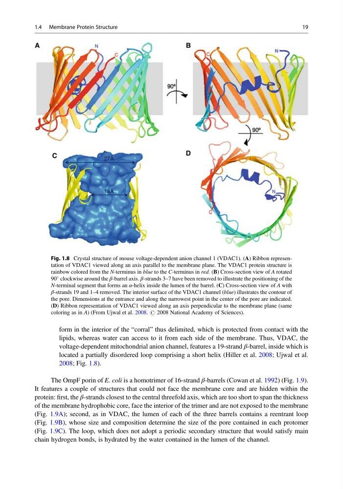
voltage-dependent mitochondrial anion channel, features a 19-strand β-barrel, inside which is
located a partially disordered loop comprising a short helix (Hiller et al. 2008; Ujwal et al.
2008; Fig. 1.8).
The OmpF porin of E. coli is a homotrimer of 16-strand β-barrels (Cowan et al. 1992) (Fig. 1.9).
It features a couple of structures that could not face the membrane core and are hidden within the
protein: first, the β-strands closest to the central threefold axis, which are too short to span the thickness
of the membrane hydrophobic core, face the interior of the trimer and are not exposed to the membrane
(Fig. 1.9A); second, as in VDAC, the lumen of each of the three barrels contains a reentrant loop
(Fig. 1.9B), whose size and composition determine the size of the pore contained in each protomer
(Fig. 1.9C). The loop, which does not adopt a periodic secondary structure that would satisfy main
chain hydrogen bonds, is hydrated by the water contained in the lumen of the channel.
Fig. 1.8 Crystal structure of mouse voltage-dependent anion channel 1 (VDAC1). (A) Ribbon representation of VDAC1 viewed along an axis parallel to the membrane plane. The VDAC1 protein structure is
rainbow colored from the N-terminus in blue to the C-terminus in red. (B) Cross-section view of A rotated
90
clockwise around the β-barrel axis. β-strands 3–7 have been removed to illustrate the positioning of the
N-terminal segment that forms an α-helix inside the lumen of the barrel. (C) Cross-section view of A with
β-strands 19 and 1–4 removed. The interior surface of the VDAC1 channel (blue) illustrates the contour of
the pore. Dimensions at the entrance and along the narrowest point in the center of the pore are indicated.
(D) Ribbon representation of VDAC1 viewed along an axis perpendicular to the membrane plane (same
coloring as in A) (From Ujwal et al. 2008. # 2008 National Academy of Sciences).
1.4 Membrane Protein Structure
19
lipids, whereas water can access to it from each side of the membrane. Thus, VDAC, the
voltage-dependent mitochondrial anion channel, features a 19-strand β-barrel, inside which is
located a partially disordered loop comprising a short helix (Hiller et al. 2008; Ujwal et al.
2008; Fig. 1.8).
The OmpF porin of E. coli is a homotrimer of 16-strand β-barrels (Cowan et al. 1992) (Fig. 1.9).
It features a couple of structures that could not face the membrane core and are hidden within the
protein: first, the β-strands closest to the central threefold axis, which are too short to span the thickness
of the membrane hydrophobic core, face the interior of the trimer and are not exposed to the membrane
(Fig. 1.9A); second, as in VDAC, the lumen of each of the three barrels contains a reentrant loop
(Fig. 1.9B), whose size and composition determine the size of the pore contained in each protomer
(Fig. 1.9C). The loop, which does not adopt a periodic secondary structure that would satisfy main
chain hydrogen bonds, is hydrated by the water contained in the lumen of the channel.
Fig. 1.8 Crystal structure of mouse voltage-dependent anion channel 1 (VDAC1). (A) Ribbon representation of VDAC1 viewed along an axis parallel to the membrane plane. The VDAC1 protein structure is
rainbow colored from the N-terminus in blue to the C-terminus in red. (B) Cross-section view of A rotated
90
clockwise around the β-barrel axis. β-strands 3–7 have been removed to illustrate the positioning of the
N-terminal segment that forms an α-helix inside the lumen of the barrel. (C) Cross-section view of A with
β-strands 19 and 1–4 removed. The interior surface of the VDAC1 channel (blue) illustrates the contour of
the pore. Dimensions at the entrance and along the narrowest point in the center of the pore are indicated.
(D) Ribbon representation of VDAC1 viewed along an axis perpendicular to the membrane plane (same
coloring as in A) (From Ujwal et al. 2008. # 2008 National Academy of Sciences).
1.4 Membrane Protein Structure
19