

2. A single α-helix, provided its central region is mostly hydrophobic, satisfies the above
conditions, and many MPs feature only one, two, or three TM helices (Figs. 1.5B and 1.6).
Such proteins can be monomeric, or assemble into homo- or hetero-oligomers. MPs whose
TM segments are made of β-strands, on the contrary, must feature enough of them – that is, at
least eight – to form a β-sheet which will close upon itself into a β-barrel, so that its TM edges
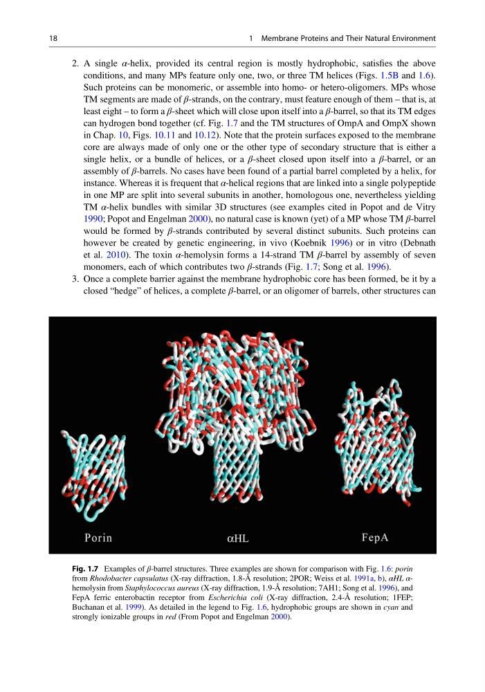
can hydrogen bond together (cf. Fig. 1.7 and the TM structures of OmpA and OmpX shown
in Chap. 10, Figs. 10.11 and 10.12). Note that the protein surfaces exposed to the membrane
core are always made of only one or the other type of secondary structure that is either a
single helix, or a bundle of helices, or a β-sheet closed upon itself into a β-barrel, or an
assembly of β-barrels. No cases have been found of a partial barrel completed by a helix, for
instance. Whereas it is frequent that α-helical regions that are linked into a single polypeptide
in one MP are split into several subunits in another, homologous one, nevertheless yielding
TM α-helix bundles with similar 3D structures (see examples cited in Popot and de Vitry
1990; Popot and Engelman 2000), no natural case is known (yet) of a MP whose TM β-barrel
would be formed by β-strands contributed by several distinct subunits. Such proteins can
however be created by genetic engineering, in vivo (Koebnik 1996) or in vitro (Debnath
et al. 2010). The toxin α-hemolysin forms a 14-strand TM β-barrel by assembly of seven
monomers, each of which contributes two β-strands (Fig. 1.7; Song et al. 1996).
3. Once a complete barrier against the membrane hydrophobic core has been formed, be it by a
closed “hedge” of helices, a complete β-barrel, or an oligomer of barrels, other structures can
Fig. 1.7 Examples of β-barrel structures. Three examples are shown for comparison with Fig. 1.6: porin
from Rhodobacter capsulatus (X-ray diffraction, 1.8-Å resolution; 2POR; Weiss et al. 1991a, b), αHL αhemolysin from Staphylococcus aureus (X-ray diffraction, 1.9-Å resolution; 7AH1; Song et al. 1996), and
FepA ferric enterobactin receptor from Escherichia coli (X-ray diffraction, 2.4-Å resolution; 1FEP;
Buchanan et al. 1999). As detailed in the legend to Fig. 1.6, hydrophobic groups are shown in cyan and
strongly ionizable groups in red (From Popot and Engelman 2000).
18
1 Membrane Proteins and Their Natural Environment
conditions, and many MPs feature only one, two, or three TM helices (Figs. 1.5B and 1.6).
Such proteins can be monomeric, or assemble into homo- or hetero-oligomers. MPs whose
TM segments are made of β-strands, on the contrary, must feature enough of them – that is, at
least eight – to form a β-sheet which will close upon itself into a β-barrel, so that its TM edges
can hydrogen bond together (cf. Fig. 1.7 and the TM structures of OmpA and OmpX shown
in Chap. 10, Figs. 10.11 and 10.12). Note that the protein surfaces exposed to the membrane
core are always made of only one or the other type of secondary structure that is either a
single helix, or a bundle of helices, or a β-sheet closed upon itself into a β-barrel, or an
assembly of β-barrels. No cases have been found of a partial barrel completed by a helix, for
instance. Whereas it is frequent that α-helical regions that are linked into a single polypeptide
in one MP are split into several subunits in another, homologous one, nevertheless yielding
TM α-helix bundles with similar 3D structures (see examples cited in Popot and de Vitry
1990; Popot and Engelman 2000), no natural case is known (yet) of a MP whose TM β-barrel
would be formed by β-strands contributed by several distinct subunits. Such proteins can
however be created by genetic engineering, in vivo (Koebnik 1996) or in vitro (Debnath
et al. 2010). The toxin α-hemolysin forms a 14-strand TM β-barrel by assembly of seven
monomers, each of which contributes two β-strands (Fig. 1.7; Song et al. 1996).
3. Once a complete barrier against the membrane hydrophobic core has been formed, be it by a
closed “hedge” of helices, a complete β-barrel, or an oligomer of barrels, other structures can
Fig. 1.7 Examples of β-barrel structures. Three examples are shown for comparison with Fig. 1.6: porin
from Rhodobacter capsulatus (X-ray diffraction, 1.8-Å resolution; 2POR; Weiss et al. 1991a, b), αHL αhemolysin from Staphylococcus aureus (X-ray diffraction, 1.9-Å resolution; 7AH1; Song et al. 1996), and
FepA ferric enterobactin receptor from Escherichia coli (X-ray diffraction, 2.4-Å resolution; 1FEP;
Buchanan et al. 1999). As detailed in the legend to Fig. 1.6, hydrophobic groups are shown in cyan and
strongly ionizable groups in red (From Popot and Engelman 2000).
18
1 Membrane Proteins and Their Natural Environment