

GPCRs expressed in the absence of surfactant, solubilized with LMPG, and transferred to NVoy).
They were found to comprise monomers and dimers in variable proportions depending on the mode of
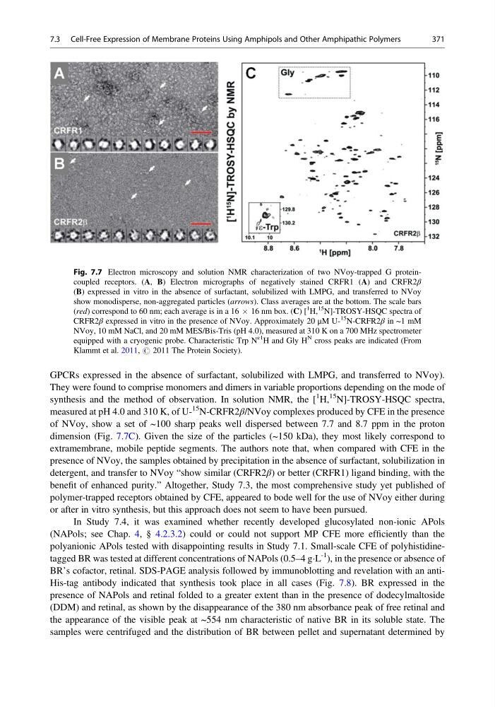
synthesis and the method of observation. In solution NMR, the [
1 H,
15 N]-TROSY-HSQC spectra,
measured at pH 4.0 and 310 K, of U15 N-CRFR2β/NVoy complexes produced by CFE in the presence
of NVoy, show a set of ~100 sharp peaks well dispersed between 7.7 and 8.7 ppm in the proton
dimension (Fig. 7.7C). Given the size of the particles (~150 kDa), they most likely correspond to
extramembrane, mobile peptide segments. The authors note that, when compared with CFE in the
presence of NVoy, the samples obtained by precipitation in the absence of surfactant, solubilization in
detergent, and transfer to NVoy “show similar (CRFR2β) or better (CRFR1) ligand binding, with the
benefit of enhanced purity.” Altogether, Study 7.3, the most comprehensive study yet published of
polymer-trapped receptors obtained by CFE, appeared to bode well for the use of NVoy either during
or after in vitro synthesis, but this approach does not seem to have been pursued.
In Study 7.4, it was examined whether recently developed glucosylated non-ionic APols
(NAPols; see Chap. 4, § 4.2.3.2) could or could not support MP CFE more efficiently than the
polyanionic APols tested with disappointing results in Study 7.1. Small-scale CFE of polyhistidinetagged BR was tested at different concentrations of NAPols (0.5–4 gÁL
-1
), in the presence or absence of
BR’s cofactor, retinal. SDS-PAGE analysis followed by immunoblotting and revelation with an antiHis-tag antibody indicated that synthesis took place in all cases (Fig. 7.8). BR expressed in the
presence of NAPols and retinal folded to a greater extent than in the presence of dodecylmaltoside
(DDM) and retinal, as shown by the disappearance of the 380 nm absorbance peak of free retinal and
the appearance of the visible peak at ~554 nm characteristic of native BR in its soluble state. The
samples were centrifuged and the distribution of BR between pellet and supernatant determined by
Fig. 7.7 Electron microscopy and solution NMR characterization of two NVoy-trapped G proteincoupled receptors. (A, B) Electron micrographs of negatively stained CRFR1 (A) and CRFR2β
(B) expressed in vitro in the absence of surfactant, solubilized with LMPG, and transferred to NVoy
show monodisperse, non-aggregated particles (arrows). Class averages are at the bottom. The scale bars
(red) correspond to 60 nm; each average is in a 16 Â 16 nm box. (C) [
1
H,
15
N]-TROSY-HSQC spectra of
CRFR2β expressed in vitro in the presence of NVoy. Approximately 20 μM U15
N-CRFR2β in ~1 mM
NVoy, 10 mM NaCl, and 20 mM MES/Bis-Tris (pH 4.0), measured at 310 K on a 700 MHz spectrometer
equipped with a cryogenic probe. Characteristic Trp N
ε1
H and Gly H
N cross peaks are indicated (From
Klammt et al. 2011, # 2011 The Protein Society).
7.3 Cell-Free Expression of Membrane Proteins Using Amphipols and Other Amphipathic Polymers
371
They were found to comprise monomers and dimers in variable proportions depending on the mode of
synthesis and the method of observation. In solution NMR, the [
1 H,
15 N]-TROSY-HSQC spectra,
measured at pH 4.0 and 310 K, of U15 N-CRFR2β/NVoy complexes produced by CFE in the presence
of NVoy, show a set of ~100 sharp peaks well dispersed between 7.7 and 8.7 ppm in the proton
dimension (Fig. 7.7C). Given the size of the particles (~150 kDa), they most likely correspond to
extramembrane, mobile peptide segments. The authors note that, when compared with CFE in the
presence of NVoy, the samples obtained by precipitation in the absence of surfactant, solubilization in
detergent, and transfer to NVoy “show similar (CRFR2β) or better (CRFR1) ligand binding, with the
benefit of enhanced purity.” Altogether, Study 7.3, the most comprehensive study yet published of
polymer-trapped receptors obtained by CFE, appeared to bode well for the use of NVoy either during
or after in vitro synthesis, but this approach does not seem to have been pursued.
In Study 7.4, it was examined whether recently developed glucosylated non-ionic APols
(NAPols; see Chap. 4, § 4.2.3.2) could or could not support MP CFE more efficiently than the
polyanionic APols tested with disappointing results in Study 7.1. Small-scale CFE of polyhistidinetagged BR was tested at different concentrations of NAPols (0.5–4 gÁL
-1
), in the presence or absence of
BR’s cofactor, retinal. SDS-PAGE analysis followed by immunoblotting and revelation with an antiHis-tag antibody indicated that synthesis took place in all cases (Fig. 7.8). BR expressed in the
presence of NAPols and retinal folded to a greater extent than in the presence of dodecylmaltoside
(DDM) and retinal, as shown by the disappearance of the 380 nm absorbance peak of free retinal and
the appearance of the visible peak at ~554 nm characteristic of native BR in its soluble state. The
samples were centrifuged and the distribution of BR between pellet and supernatant determined by
Fig. 7.7 Electron microscopy and solution NMR characterization of two NVoy-trapped G proteincoupled receptors. (A, B) Electron micrographs of negatively stained CRFR1 (A) and CRFR2β
(B) expressed in vitro in the absence of surfactant, solubilized with LMPG, and transferred to NVoy
show monodisperse, non-aggregated particles (arrows). Class averages are at the bottom. The scale bars
(red) correspond to 60 nm; each average is in a 16 Â 16 nm box. (C) [
1
H,
15
N]-TROSY-HSQC spectra of
CRFR2β expressed in vitro in the presence of NVoy. Approximately 20 μM U15
N-CRFR2β in ~1 mM
NVoy, 10 mM NaCl, and 20 mM MES/Bis-Tris (pH 4.0), measured at 310 K on a 700 MHz spectrometer
equipped with a cryogenic probe. Characteristic Trp N
ε1
H and Gly H
N cross peaks are indicated (From
Klammt et al. 2011, # 2011 The Protein Society).
7.3 Cell-Free Expression of Membrane Proteins Using Amphipols and Other Amphipathic Polymers
371