

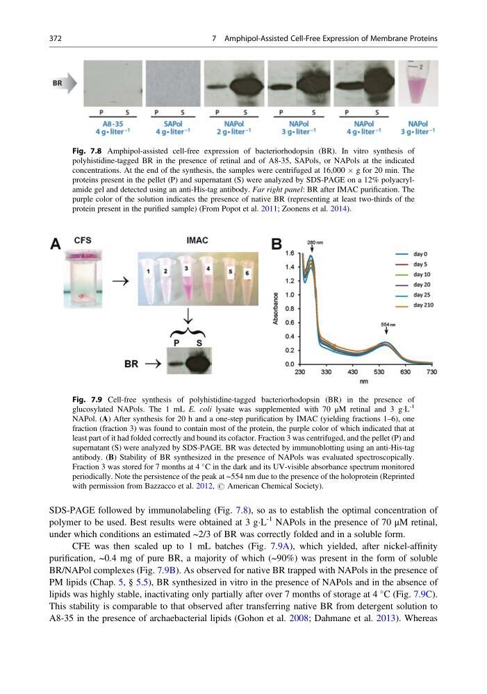
SDS-PAGE followed by immunolabeling (Fig. 7.8), so as to establish the optimal concentration of
polymer to be used. Best results were obtained at 3 gÁL
-1 NAPols in the presence of 70 μM retinal,
under which conditions an estimated ~2/3 of BR was correctly folded and in a soluble form.
CFE was then scaled up to 1 mL batches (Fig. 7.9A), which yielded, after nickel-affinity
purification, ~0.4 mg of pure BR, a majority of which (~90%) was present in the form of soluble
BR/NAPol complexes (Fig. 7.9B). As observed for native BR trapped with NAPols in the presence of
PM lipids (Chap. 5, § 5.5), BR synthesized in vitro in the presence of NAPols and in the absence of
lipids was highly stable, inactivating only partially after over 7 months of storage at 4
C (Fig. 7.9C).
This stability is comparable to that observed after transferring native BR from detergent solution to
A8-35 in the presence of archaebacterial lipids (Gohon et al. 2008; Dahmane et al. 2013). Whereas
Fig. 7.9 Cell-free synthesis of polyhistidine-tagged bacteriorhodopsin (BR) in the presence of
glucosylated NAPols. The 1 mL E. coli lysate was supplemented with 70 μM retinal and 3 gÁL
-1
NAPol. (A) After synthesis for 20 h and a one-step purification by IMAC (yielding fractions 1–6), one
fraction (fraction 3) was found to contain most of the protein, the purple color of which indicated that at
least part of it had folded correctly and bound its cofactor. Fraction 3 was centrifuged, and the pellet (P) and
supernatant (S) were analyzed by SDS-PAGE. BR was detected by immunoblotting using an anti-His-tag
antibody. (B) Stability of BR synthesized in the presence of NAPols was evaluated spectroscopically.
Fraction 3 was stored for 7 months at 4
C in the dark and its UV-visible absorbance spectrum monitored
periodically. Note the persistence of the peak at ~554 nm due to the presence of the holoprotein (Reprinted
with permission from Bazzacco et al. 2012, # American Chemical Society).
Fig. 7.8 Amphipol-assisted cell-free expression of bacteriorhodopsin (BR). In vitro synthesis of
polyhistidine-tagged BR in the presence of retinal and of A8-35, SAPols, or NAPols at the indicated
concentrations. At the end of the synthesis, the samples were centrifuged at 16,000 Â g for 20 min. The
proteins present in the pellet (P) and supernatant (S) were analyzed by SDS-PAGE on a 12% polyacrylamide gel and detected using an anti-His-tag antibody. Far right panel: BR after IMAC purification. The
purple color of the solution indicates the presence of native BR (representing at least two-thirds of the
protein present in the purified sample) (From Popot et al. 2011; Zoonens et al. 2014).
372
7 Amphipol-Assisted Cell-Free Expression of Membrane Proteins
polymer to be used. Best results were obtained at 3 gÁL
-1 NAPols in the presence of 70 μM retinal,
under which conditions an estimated ~2/3 of BR was correctly folded and in a soluble form.
CFE was then scaled up to 1 mL batches (Fig. 7.9A), which yielded, after nickel-affinity
purification, ~0.4 mg of pure BR, a majority of which (~90%) was present in the form of soluble
BR/NAPol complexes (Fig. 7.9B). As observed for native BR trapped with NAPols in the presence of
PM lipids (Chap. 5, § 5.5), BR synthesized in vitro in the presence of NAPols and in the absence of
lipids was highly stable, inactivating only partially after over 7 months of storage at 4
C (Fig. 7.9C).
This stability is comparable to that observed after transferring native BR from detergent solution to
A8-35 in the presence of archaebacterial lipids (Gohon et al. 2008; Dahmane et al. 2013). Whereas
Fig. 7.9 Cell-free synthesis of polyhistidine-tagged bacteriorhodopsin (BR) in the presence of
glucosylated NAPols. The 1 mL E. coli lysate was supplemented with 70 μM retinal and 3 gÁL
-1
NAPol. (A) After synthesis for 20 h and a one-step purification by IMAC (yielding fractions 1–6), one
fraction (fraction 3) was found to contain most of the protein, the purple color of which indicated that at
least part of it had folded correctly and bound its cofactor. Fraction 3 was centrifuged, and the pellet (P) and
supernatant (S) were analyzed by SDS-PAGE. BR was detected by immunoblotting using an anti-His-tag
antibody. (B) Stability of BR synthesized in the presence of NAPols was evaluated spectroscopically.
Fraction 3 was stored for 7 months at 4
C in the dark and its UV-visible absorbance spectrum monitored
periodically. Note the persistence of the peak at ~554 nm due to the presence of the holoprotein (Reprinted
with permission from Bazzacco et al. 2012, # American Chemical Society).
Fig. 7.8 Amphipol-assisted cell-free expression of bacteriorhodopsin (BR). In vitro synthesis of
polyhistidine-tagged BR in the presence of retinal and of A8-35, SAPols, or NAPols at the indicated
concentrations. At the end of the synthesis, the samples were centrifuged at 16,000 Â g for 20 min. The
proteins present in the pellet (P) and supernatant (S) were analyzed by SDS-PAGE on a 12% polyacrylamide gel and detected using an anti-His-tag antibody. Far right panel: BR after IMAC purification. The
purple color of the solution indicates the presence of native BR (representing at least two-thirds of the
protein present in the purified sample) (From Popot et al. 2011; Zoonens et al. 2014).
372
7 Amphipol-Assisted Cell-Free Expression of Membrane Proteins