

non-specifically to A8-35 particles. This causes difficulties in assessing by ligand binding the extent of
folding achieved in A8-35. Measurements are easier when the ghrelin receptor is folded in NAPols,
where the background is lower. Ligand-binding measurements indicated a folding yield of ~40%, and
a receptor ~97% active was obtained after affinity chromatography (Bazzacco et al. 2012). The binding
properties of the folded GHSR-1a were then further examined to assess the quality of folding.
Fluorescence energy transfer from GHSR-1a, labeled with Alexa Fluor 350, to a ghrelin peptide
labeled with fluorescein isothiocyanate was recorded in competition experiments with synthetic
antagonists. The competition profiles obtained by this method are within the same range as those
previously inferred from radioactive and TagLite-based measurements on human embryonic kidney
(HEK) cells transiently expressing GHSR-1a (Leyris et al. 2011). In addition, GHSR-1a folded in
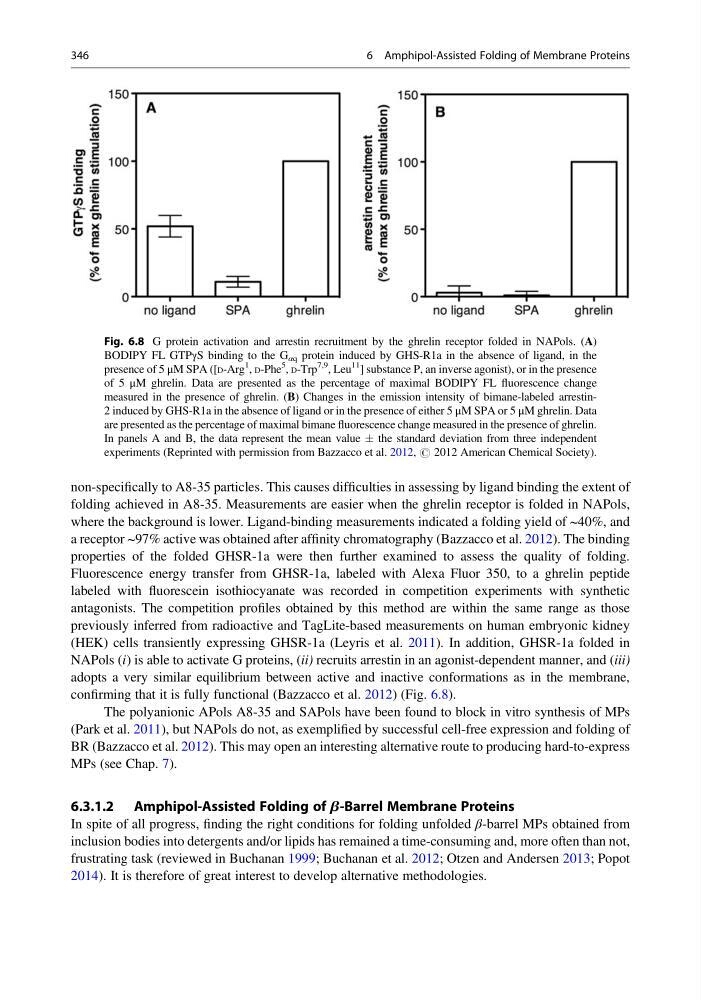
NAPols (i) is able to activate G proteins, (ii) recruits arrestin in an agonist-dependent manner, and (iii)
adopts a very similar equilibrium between active and inactive conformations as in the membrane,
confirming that it is fully functional (Bazzacco et al. 2012) (Fig. 6.8).
The polyanionic APols A8-35 and SAPols have been found to block in vitro synthesis of MPs
(Park et al. 2011), but NAPols do not, as exemplified by successful cell-free expression and folding of
BR (Bazzacco et al. 2012). This may open an interesting alternative route to producing hard-to-express
MPs (see Chap. 7).
6.3.1.2 Amphipol-Assisted Folding of b-Barrel Membrane Proteins
In spite of all progress, finding the right conditions for folding unfolded β-barrel MPs obtained from
inclusion bodies into detergents and/or lipids has remained a time-consuming and, more often than not,
frustrating task (reviewed in Buchanan 1999; Buchanan et al. 2012; Otzen and Andersen 2013; Popot
2014). It is therefore of great interest to develop alternative methodologies.
Fig. 6.8 G protein activation and arrestin recruitment by the ghrelin receptor folded in NAPols. (A)
BODIPY FL GTPγS binding to the G αq protein induced by GHS-R1a in the absence of ligand, in the
presence of 5 μM SPA ([D-Arg
1
, D-Phe
5
, D-Trp
7,9
, Leu
11
] substance P, an inverse agonist), or in the presence
of 5 μM ghrelin. Data are presented as the percentage of maximal BODIPY FL fluorescence change
measured in the presence of ghrelin. (B) Changes in the emission intensity of bimane-labeled arrestin2 induced by GHS-R1a in the absence of ligand or in the presence of either 5 μM SPA or 5 μM ghrelin. Data
are presented as the percentage of maximal bimane fluorescence change measured in the presence of ghrelin.
In panels A and B, the data represent the mean value Æ the standard deviation from three independent
experiments (Reprinted with permission from Bazzacco et al. 2012, # 2012 American Chemical Society).
346
6 Amphipol-Assisted Folding of Membrane Proteins
folding achieved in A8-35. Measurements are easier when the ghrelin receptor is folded in NAPols,
where the background is lower. Ligand-binding measurements indicated a folding yield of ~40%, and
a receptor ~97% active was obtained after affinity chromatography (Bazzacco et al. 2012). The binding
properties of the folded GHSR-1a were then further examined to assess the quality of folding.
Fluorescence energy transfer from GHSR-1a, labeled with Alexa Fluor 350, to a ghrelin peptide
labeled with fluorescein isothiocyanate was recorded in competition experiments with synthetic
antagonists. The competition profiles obtained by this method are within the same range as those
previously inferred from radioactive and TagLite-based measurements on human embryonic kidney
(HEK) cells transiently expressing GHSR-1a (Leyris et al. 2011). In addition, GHSR-1a folded in
NAPols (i) is able to activate G proteins, (ii) recruits arrestin in an agonist-dependent manner, and (iii)
adopts a very similar equilibrium between active and inactive conformations as in the membrane,
confirming that it is fully functional (Bazzacco et al. 2012) (Fig. 6.8).
The polyanionic APols A8-35 and SAPols have been found to block in vitro synthesis of MPs
(Park et al. 2011), but NAPols do not, as exemplified by successful cell-free expression and folding of
BR (Bazzacco et al. 2012). This may open an interesting alternative route to producing hard-to-express
MPs (see Chap. 7).
6.3.1.2 Amphipol-Assisted Folding of b-Barrel Membrane Proteins
In spite of all progress, finding the right conditions for folding unfolded β-barrel MPs obtained from
inclusion bodies into detergents and/or lipids has remained a time-consuming and, more often than not,
frustrating task (reviewed in Buchanan 1999; Buchanan et al. 2012; Otzen and Andersen 2013; Popot
2014). It is therefore of great interest to develop alternative methodologies.
Fig. 6.8 G protein activation and arrestin recruitment by the ghrelin receptor folded in NAPols. (A)
BODIPY FL GTPγS binding to the G αq protein induced by GHS-R1a in the absence of ligand, in the
presence of 5 μM SPA ([D-Arg
1
, D-Phe
5
, D-Trp
7,9
, Leu
11
] substance P, an inverse agonist), or in the presence
of 5 μM ghrelin. Data are presented as the percentage of maximal BODIPY FL fluorescence change
measured in the presence of ghrelin. (B) Changes in the emission intensity of bimane-labeled arrestin2 induced by GHS-R1a in the absence of ligand or in the presence of either 5 μM SPA or 5 μM ghrelin. Data
are presented as the percentage of maximal bimane fluorescence change measured in the presence of ghrelin.
In panels A and B, the data represent the mean value Æ the standard deviation from three independent
experiments (Reprinted with permission from Bazzacco et al. 2012, # 2012 American Chemical Society).
346
6 Amphipol-Assisted Folding of Membrane Proteins