

6.3.1.2.1 Folding of OmpA and FomA in A8-35
Applying APols to (re)folding outer membrane proteins was first attempted using as models OmpA
from E. coli and FomA from Fusobacterium nucleatum, two monomeric β-barrels with, respectively,
8 (observed) and 14 (predicted) TM β-strands. OmpA features, in addition, a periplasmic C-terminal
domain (~17 kDa), which can be genetically removed, yielding the isolated TM domain (tOmpA).
Folding was achieved according to the very simple scheme shown in Fig. 6.5B: The proteins,
expressed as inclusion bodies, are dissolved in unfolded form in either 8 M (OmpA) or 10 M urea
(FomA), and the preparations diluted ~20-fold in a solution of A8-35 (Pocanschi et al. 2006). Folding
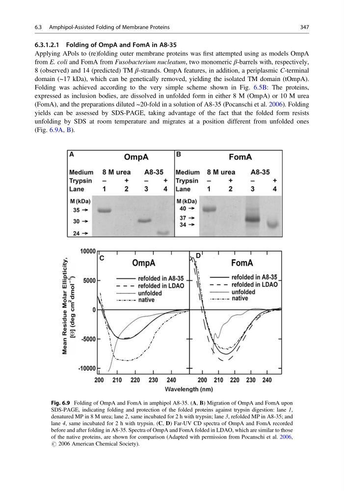
yields can be assessed by SDS-PAGE, taking advantage of the fact that the folded form resists
unfolding by SDS at room temperature and migrates at a position different from unfolded ones
(Fig. 6.9A, B).
Fig. 6.9 Folding of OmpA and FomA in amphipol A8-35. (A, B) Migration of OmpA and FomA upon
SDS-PAGE, indicating folding and protection of the folded proteins against trypsin digestion: lane 1,
denatured MP in 8 M urea; lane 2, same incubated for 2 h with trypsin; lane 3, refolded MP in A8-35; and
lane 4, same incubated for 2 h with trypsin. (C, D) Far-UV CD spectra of OmpA and FomA recorded
before and after folding in A8-35. Spectra of OmpA and FomA folded in LDAO, which are similar to those
of the native proteins, are shown for comparison (Adapted with permission from Pocanschi et al. 2006,
# 2006 American Chemical Society).
6.3 Amphipol-Assisted Folding of Membrane Proteins
347
Applying APols to (re)folding outer membrane proteins was first attempted using as models OmpA
from E. coli and FomA from Fusobacterium nucleatum, two monomeric β-barrels with, respectively,
8 (observed) and 14 (predicted) TM β-strands. OmpA features, in addition, a periplasmic C-terminal
domain (~17 kDa), which can be genetically removed, yielding the isolated TM domain (tOmpA).
Folding was achieved according to the very simple scheme shown in Fig. 6.5B: The proteins,
expressed as inclusion bodies, are dissolved in unfolded form in either 8 M (OmpA) or 10 M urea
(FomA), and the preparations diluted ~20-fold in a solution of A8-35 (Pocanschi et al. 2006). Folding
yields can be assessed by SDS-PAGE, taking advantage of the fact that the folded form resists
unfolding by SDS at room temperature and migrates at a position different from unfolded ones
(Fig. 6.9A, B).
Fig. 6.9 Folding of OmpA and FomA in amphipol A8-35. (A, B) Migration of OmpA and FomA upon
SDS-PAGE, indicating folding and protection of the folded proteins against trypsin digestion: lane 1,
denatured MP in 8 M urea; lane 2, same incubated for 2 h with trypsin; lane 3, refolded MP in A8-35; and
lane 4, same incubated for 2 h with trypsin. (C, D) Far-UV CD spectra of OmpA and FomA recorded
before and after folding in A8-35. Spectra of OmpA and FomA folded in LDAO, which are similar to those
of the native proteins, are shown for comparison (Adapted with permission from Pocanschi et al. 2006,
# 2006 American Chemical Society).
6.3 Amphipol-Assisted Folding of Membrane Proteins
347