

It is probably relevant to this issue that the detergent-induced denaturation of SERCA1a seems to
start with the opening of the transmembrane helix bundle, with the Ca
2+ -binding TM region being
unfolded first (Merino et al. 1994). The fact that ATPase inactivation in the presence of detergent is
faster in the absence of Ca
2+ than in its presence is probably related, at least in part, to the “crosslinking” of the TM helices by two Ca
2+ ions, which bind to negatively charged residues in TM helices
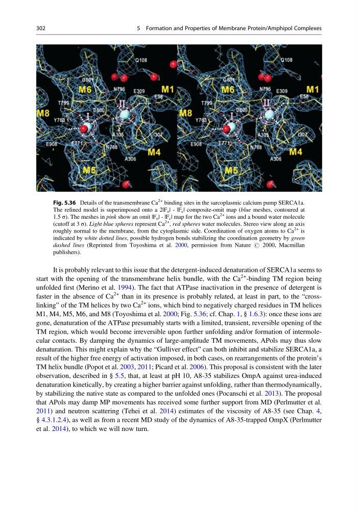
M1, M4, M5, M6, and M8 (Toyoshima et al. 2000; Fig. 5.36; cf. Chap. 1, § 1.6.3): once these ions are
gone, denaturation of the ATPase presumably starts with a limited, transient, reversible opening of the
TM region, which would become irreversible upon further unfolding and/or formation of intermolecular contacts. By damping the dynamics of large-amplitude TM movements, APols may thus slow
denaturation. This might explain why the “Gulliver effect” can both inhibit and stabilize SERCA1a, a
result of the higher free energy of activation imposed, in both cases, on rearrangements of the protein’s
TM helix bundle (Popot et al. 2003, 2011; Picard et al. 2006). This proposal is consistent with the later
observation, described in § 5.5, that, at least at pH 10, A8-35 stabilizes OmpA against urea-induced
denaturation kinetically, by creating a higher barrier against unfolding, rather than thermodynamically,
by stabilizing the native state as compared to the unfolded ones (Pocanschi et al. 2013). The proposal
that APols may damp MP movements has received some further support from MD (Perlmutter et al.
2011) and neutron scattering (Tehei et al. 2014) estimates of the viscosity of A8-35 (see Chap. 4,
§ 4.3.1.2.4), as well as from a recent MD study of the dynamics of A8-35-trapped OmpX (Perlmutter
et al. 2014), to which we will now turn.
Fig. 5.36 Details of the transmembrane Ca
2+ binding sites in the sarcoplasmic calcium pump SERCA1a.
The refined model is superimposed onto a 2|F o | - |F c | composite-omit map (blue meshes, contoured at
1.5 σ). The meshes in pink show an omit |F o | - |F c | map for the two Ca
2+ ions and a bound water molecule
(cutoff at 3 σ). Light blue spheres represent Ca
2+ , red spheres water molecules. Stereo view along an axis
roughly normal to the membrane, from the cytoplasmic side. Coordination of oxygen atoms to Ca
2+ is
indicated by white dotted lines, possible hydrogen bonds stabilizing the coordination geometry by green
dashed lines (Reprinted from Toyoshima et al. 2000, permission from Nature # 2000, Macmillan
publishers).
302
5 Formation and Properties of Membrane Protein/Amphipol Complexes
start with the opening of the transmembrane helix bundle, with the Ca
2+ -binding TM region being
unfolded first (Merino et al. 1994). The fact that ATPase inactivation in the presence of detergent is
faster in the absence of Ca
2+ than in its presence is probably related, at least in part, to the “crosslinking” of the TM helices by two Ca
2+ ions, which bind to negatively charged residues in TM helices
M1, M4, M5, M6, and M8 (Toyoshima et al. 2000; Fig. 5.36; cf. Chap. 1, § 1.6.3): once these ions are
gone, denaturation of the ATPase presumably starts with a limited, transient, reversible opening of the
TM region, which would become irreversible upon further unfolding and/or formation of intermolecular contacts. By damping the dynamics of large-amplitude TM movements, APols may thus slow
denaturation. This might explain why the “Gulliver effect” can both inhibit and stabilize SERCA1a, a
result of the higher free energy of activation imposed, in both cases, on rearrangements of the protein’s
TM helix bundle (Popot et al. 2003, 2011; Picard et al. 2006). This proposal is consistent with the later
observation, described in § 5.5, that, at least at pH 10, A8-35 stabilizes OmpA against urea-induced
denaturation kinetically, by creating a higher barrier against unfolding, rather than thermodynamically,
by stabilizing the native state as compared to the unfolded ones (Pocanschi et al. 2013). The proposal
that APols may damp MP movements has received some further support from MD (Perlmutter et al.
2011) and neutron scattering (Tehei et al. 2014) estimates of the viscosity of A8-35 (see Chap. 4,
§ 4.3.1.2.4), as well as from a recent MD study of the dynamics of A8-35-trapped OmpX (Perlmutter
et al. 2014), to which we will now turn.
Fig. 5.36 Details of the transmembrane Ca
2+ binding sites in the sarcoplasmic calcium pump SERCA1a.
The refined model is superimposed onto a 2|F o | - |F c | composite-omit map (blue meshes, contoured at
1.5 σ). The meshes in pink show an omit |F o | - |F c | map for the two Ca
2+ ions and a bound water molecule
(cutoff at 3 σ). Light blue spheres represent Ca
2+ , red spheres water molecules. Stereo view along an axis
roughly normal to the membrane, from the cytoplasmic side. Coordination of oxygen atoms to Ca
2+ is
indicated by white dotted lines, possible hydrogen bonds stabilizing the coordination geometry by green
dashed lines (Reprinted from Toyoshima et al. 2000, permission from Nature # 2000, Macmillan
publishers).
302
5 Formation and Properties of Membrane Protein/Amphipol Complexes