

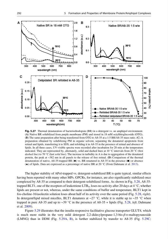
The higher stability of APol-trapped vs. detergent-solubilized BR is quite typical, similar effects
having been reported with many other MPs. GPCRs, for instance, are also significantly stabilized once
complexed by A8-35 as compared to their detergent-solubilized forms. As shown in Fig. 5.28, A8-35trapped BLT1, one of the receptors of leukotriene LTB 4 , loses no activity after 20 days at 4
C, whether
lipids are present or not, whereas, under the same conditions of buffer and temperature, BLT1 kept in
fos-choline-16/asolectin solution loses about half of its activity over the same period (Fig. 5.28, right).
In detergent/lipid mixed micelles, BLT1 denatures at ~27
C, while it is stable up to ~35
C when
trapped in pure A8-35 and up to ~39
C in the presence of A8-35 + lipids (Fig. 5.28, left; Dahmane
et al. 2009).
Figure 5.29 illustrates that the insulin-responsive facilitative glucose transporter GLUT4, which
is much more stable in the very mild detergent 2,2-didecylpropane-1,3-bis-β-D-maltopyranoside
(LMNG) than in DDM (Fig. 5.29A, B), is further stabilized by transfer to A8-35 (Fig. 5.29C;
Fig. 5.27 Thermal denaturation of bacteriorhodopsin (BR) in a detergent vs. an amphipol environment.
(A) Native BR solubilized from purple membrane (PM) and stored in 18 mM octylthioglucoside (OTG).
(B) The same preparation after being transferred from OTG to A8-35 at a 1:5 BR/A8-35 mass ratio. (C) A
preparation obtained by solubilizing PM in organic solvent, separating the denatured apoprotein from
retinal and lipids, transferring it to SDS, and refolding it in A8-35 in the presence of retinal and absence of
lipids. In all three cases, UV-visible spectra were recorded after incubation for 20 min at the temperature
indicated. They are represented by, alternately, solid and dashed lines at 10
C intervals from 20
C (first
dashed line) to 70
C (last solid line). The increase in turbidity in A is due to aggregation of the denatured
protein, the peak at ~382 nm in all panels to the release of free retinal. (D) Comparison of the thermal
denaturation of native, A8-35-trapped BR (●) vs. BR renatured in A8-35 in the presence (■) or absence
(~) of lipids. Data are expressed as a percentage of native BR at 20
C (From Dahmane et al. 2013).
292
5 Formation and Properties of Membrane Protein/Amphipol Complexes
having been reported with many other MPs. GPCRs, for instance, are also significantly stabilized once
complexed by A8-35 as compared to their detergent-solubilized forms. As shown in Fig. 5.28, A8-35trapped BLT1, one of the receptors of leukotriene LTB 4 , loses no activity after 20 days at 4
C, whether
lipids are present or not, whereas, under the same conditions of buffer and temperature, BLT1 kept in
fos-choline-16/asolectin solution loses about half of its activity over the same period (Fig. 5.28, right).
In detergent/lipid mixed micelles, BLT1 denatures at ~27
C, while it is stable up to ~35
C when
trapped in pure A8-35 and up to ~39
C in the presence of A8-35 + lipids (Fig. 5.28, left; Dahmane
et al. 2009).
Figure 5.29 illustrates that the insulin-responsive facilitative glucose transporter GLUT4, which
is much more stable in the very mild detergent 2,2-didecylpropane-1,3-bis-β-D-maltopyranoside
(LMNG) than in DDM (Fig. 5.29A, B), is further stabilized by transfer to A8-35 (Fig. 5.29C;
Fig. 5.27 Thermal denaturation of bacteriorhodopsin (BR) in a detergent vs. an amphipol environment.
(A) Native BR solubilized from purple membrane (PM) and stored in 18 mM octylthioglucoside (OTG).
(B) The same preparation after being transferred from OTG to A8-35 at a 1:5 BR/A8-35 mass ratio. (C) A
preparation obtained by solubilizing PM in organic solvent, separating the denatured apoprotein from
retinal and lipids, transferring it to SDS, and refolding it in A8-35 in the presence of retinal and absence of
lipids. In all three cases, UV-visible spectra were recorded after incubation for 20 min at the temperature
indicated. They are represented by, alternately, solid and dashed lines at 10
C intervals from 20
C (first
dashed line) to 70
C (last solid line). The increase in turbidity in A is due to aggregation of the denatured
protein, the peak at ~382 nm in all panels to the release of free retinal. (D) Comparison of the thermal
denaturation of native, A8-35-trapped BR (●) vs. BR renatured in A8-35 in the presence (■) or absence
(~) of lipids. Data are expressed as a percentage of native BR at 20
C (From Dahmane et al. 2013).
292
5 Formation and Properties of Membrane Protein/Amphipol Complexes