

In the case of the nicotinic acetylcholine receptor (nAChR), functional data can also be
interpreted as indicating that transfer from detergent (CHAPS) to APol (A8-35) allows lipids to rebind,
but the evidence is indirect and the interpretation is not unique (Martinez et al. 2002; see § 5.4,
Fig. 5.25).
MPs are often claimed to be extracted by SMA along with a complete annulus of membrane
lipids. The “cookie cutter” simile occasionally used to describe this process is amusing, but it is
misleading, the reality being far more complex. In most studies, the amount of lipids is simply
estimated from the size of the Lipodisqs, as deduced from either EM images of negatively stained
preparations or from DLS measurements (see e.g. Long et al. 2013). In some studies, a qualitative
analysis was performed. In most cases, it shows that the lipid composition of the particles is similar to
that of the membranes the proteins were extracted from (Long et al. 2013; Dörr et al. 2014; Swainsbury
et al. 2014; cf. Fig. 5.16A, B). In the case of the SecYEG complex, however, the particles were found
to be enriched in negatively charged lipids (which are known to be important functionally) (Fig. 5.16C,
D), whereas the lipids accompanying other MPs extracted from the same E. coli membrane had the
same composition as the membrane (Prabudiansyah et al. 2015). Following expression in Sf9 insect
cells of the human equilibrative nucleoside transporter-1 (hENT1), extraction with SMA, and lipid
analysis, it was observed that polyunsaturated lipids were specifically excluded from the complexes
(Rehan et al. 2017).
In the few cases where quantitative analyses were performed, the results are case-dependent. In
the case of bacterial photosynthetic reaction centers, it was estimated that the lipids extracted along
with the protein (~150 of them) are sufficient to form three layers around it (Swainsbury et al. 2014).
The dimer of the cation diffusion facilitator CzcD was found to retain 32–35 phospholipids, which
were estimated to be able to form about 1 layer around its TM region (Bersch et al. 2017). In the case of
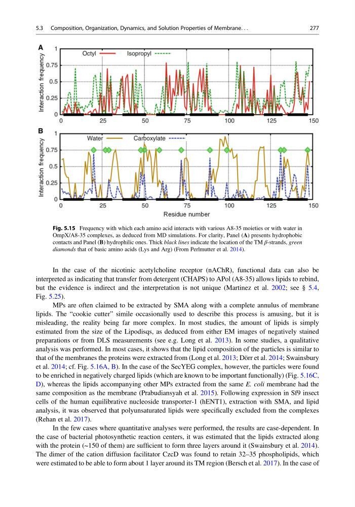
Fig. 5.15 Frequency with which each amino acid interacts with various A8-35 moieties or with water in
OmpX/A8-35 complexes, as deduced from MD simulations. For clarity, Panel (A) presents hydrophobic
contacts and Panel (B) hydrophilic ones. Thick black lines indicate the location of the TM β-strands, green
diamonds that of basic amino acids (Lys and Arg) (From Perlmutter et al. 2014).
5.3 Composition, Organization, Dynamics, and Solution Properties of Membrane. . .
277
interpreted as indicating that transfer from detergent (CHAPS) to APol (A8-35) allows lipids to rebind,
but the evidence is indirect and the interpretation is not unique (Martinez et al. 2002; see § 5.4,
Fig. 5.25).
MPs are often claimed to be extracted by SMA along with a complete annulus of membrane
lipids. The “cookie cutter” simile occasionally used to describe this process is amusing, but it is
misleading, the reality being far more complex. In most studies, the amount of lipids is simply
estimated from the size of the Lipodisqs, as deduced from either EM images of negatively stained
preparations or from DLS measurements (see e.g. Long et al. 2013). In some studies, a qualitative
analysis was performed. In most cases, it shows that the lipid composition of the particles is similar to
that of the membranes the proteins were extracted from (Long et al. 2013; Dörr et al. 2014; Swainsbury
et al. 2014; cf. Fig. 5.16A, B). In the case of the SecYEG complex, however, the particles were found
to be enriched in negatively charged lipids (which are known to be important functionally) (Fig. 5.16C,
D), whereas the lipids accompanying other MPs extracted from the same E. coli membrane had the
same composition as the membrane (Prabudiansyah et al. 2015). Following expression in Sf9 insect
cells of the human equilibrative nucleoside transporter-1 (hENT1), extraction with SMA, and lipid
analysis, it was observed that polyunsaturated lipids were specifically excluded from the complexes
(Rehan et al. 2017).
In the few cases where quantitative analyses were performed, the results are case-dependent. In
the case of bacterial photosynthetic reaction centers, it was estimated that the lipids extracted along
with the protein (~150 of them) are sufficient to form three layers around it (Swainsbury et al. 2014).
The dimer of the cation diffusion facilitator CzcD was found to retain 32–35 phospholipids, which
were estimated to be able to form about 1 layer around its TM region (Bersch et al. 2017). In the case of
Fig. 5.15 Frequency with which each amino acid interacts with various A8-35 moieties or with water in
OmpX/A8-35 complexes, as deduced from MD simulations. For clarity, Panel (A) presents hydrophobic
contacts and Panel (B) hydrophilic ones. Thick black lines indicate the location of the TM β-strands, green
diamonds that of basic amino acids (Lys and Arg) (From Perlmutter et al. 2014).
5.3 Composition, Organization, Dynamics, and Solution Properties of Membrane. . .
277