

AcrB, only ~40 lipids are retained, which was deemed insufficient to form a single layer around
the protein, implying that the latter must come into contact with the styrene groups of the polymer
(Postis et al. 2015). A similar situation prevails for SMA-trapped P-glycoprotein 1 (PgP), which is
accompanied by only ~11 lipids (Knowles et al. 2009), in keeping with cryo-EM data, which do not
show the presence of a lipid disc (Gulati et al. 2014). Similarly, hENT1/SMA complexes contain only
18 phospholipid molecules per protein (Rehan et al. 2017). The similarity between a BR/lipid/A8-35
complex, in which the protein has rebound all ten lipids it was in contact with in PM (Gohon et al.
2008), and a PgP/lipid/SMA one, in which it has retained ~11, is more striking than the difference.
Attractive as the notion may seem, distinguishing MP/A8-35 and MP/SMA complexes as non-bilayer
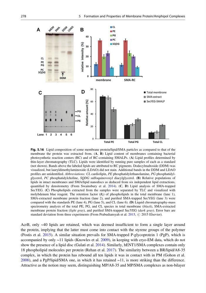
Fig. 5.16 Lipid composition of some membrane protein/lipid/SMA particles as compared to that of the
membrane the protein was extracted from. (A, B) Lipid content of membranes containing bacterial
photosynthetic reaction centers (RC) and of RC-containing SMALPs. (A) Lipid profiles determined by
thin-layer chromatography (TLC). Lipids were identified by running pure samples of each as a standard
(not shown). Bands above the labeled lipids are attributed to RC pigments. Dodecylmaltoside (DDM) was
visualized, but lauryldimethylaminoxide (LDAO) did not stain. Additional bands in the DDM and LDAO
profiles are unidentified. Abbreviations: CL cardiolipin, PE phosphatidylethanolamine, PG phosphatidylglycerol, PC phosphatidylcholine, SQDG sulfoquinovosyl diacylglycerol. (B) Relative populations of
lipids in intact membranes and SMA/lipid nanodiscs as deduced from six independent lipid extractions,
quantified by densitometry (From Swainsbury et al. 2014). (C, D) Lipid analysis of SMA-trapped
SecYEG. (C) Phospholipids extracted from the samples were separated by TLC and visualized with
molybdenum blue reagent. The retention factor (R f ) of phospholipids in the total membrane (lane 1),
SMA-extracted membrane protein fraction (lane 2), and purified SMA-trapped SecYEG (lane 3) were
compared with the standards PE (lane 4), PG (lane 5), and CL (lane 6). (D) Liquid chromatography-mass
spectrometry analysis of the total PE, PG, and CL species in total membrane (black), SMA-extracted
membrane protein fraction (light gray), and purified SMA-trapped SecYEG (dark gray). Error bars are
standard deviation from three experiments (From Prabudiansyah et al. 2015, # 2015 Elsevier).
278
5 Formation and Properties of Membrane Protein/Amphipol Complexes
the protein, implying that the latter must come into contact with the styrene groups of the polymer
(Postis et al. 2015). A similar situation prevails for SMA-trapped P-glycoprotein 1 (PgP), which is
accompanied by only ~11 lipids (Knowles et al. 2009), in keeping with cryo-EM data, which do not
show the presence of a lipid disc (Gulati et al. 2014). Similarly, hENT1/SMA complexes contain only
18 phospholipid molecules per protein (Rehan et al. 2017). The similarity between a BR/lipid/A8-35
complex, in which the protein has rebound all ten lipids it was in contact with in PM (Gohon et al.
2008), and a PgP/lipid/SMA one, in which it has retained ~11, is more striking than the difference.
Attractive as the notion may seem, distinguishing MP/A8-35 and MP/SMA complexes as non-bilayer
Fig. 5.16 Lipid composition of some membrane protein/lipid/SMA particles as compared to that of the
membrane the protein was extracted from. (A, B) Lipid content of membranes containing bacterial
photosynthetic reaction centers (RC) and of RC-containing SMALPs. (A) Lipid profiles determined by
thin-layer chromatography (TLC). Lipids were identified by running pure samples of each as a standard
(not shown). Bands above the labeled lipids are attributed to RC pigments. Dodecylmaltoside (DDM) was
visualized, but lauryldimethylaminoxide (LDAO) did not stain. Additional bands in the DDM and LDAO
profiles are unidentified. Abbreviations: CL cardiolipin, PE phosphatidylethanolamine, PG phosphatidylglycerol, PC phosphatidylcholine, SQDG sulfoquinovosyl diacylglycerol. (B) Relative populations of
lipids in intact membranes and SMA/lipid nanodiscs as deduced from six independent lipid extractions,
quantified by densitometry (From Swainsbury et al. 2014). (C, D) Lipid analysis of SMA-trapped
SecYEG. (C) Phospholipids extracted from the samples were separated by TLC and visualized with
molybdenum blue reagent. The retention factor (R f ) of phospholipids in the total membrane (lane 1),
SMA-extracted membrane protein fraction (lane 2), and purified SMA-trapped SecYEG (lane 3) were
compared with the standards PE (lane 4), PG (lane 5), and CL (lane 6). (D) Liquid chromatography-mass
spectrometry analysis of the total PE, PG, and CL species in total membrane (black), SMA-extracted
membrane protein fraction (light gray), and purified SMA-trapped SecYEG (dark gray). Error bars are
standard deviation from three experiments (From Prabudiansyah et al. 2015, # 2015 Elsevier).
278
5 Formation and Properties of Membrane Protein/Amphipol Complexes