

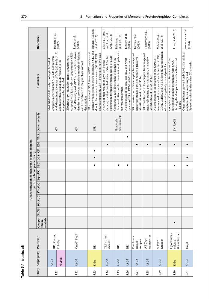
Table 5.4
(continued)
5.21
A8-35
BR, tOmpA,
b
6 f, bc
1
MS
MALDI-TOF-MS analysis of eight MP/APol
complexes confirms that APols do not interfere
with determining the mass of MPs (cf. Study 5.14)
and that protein-bound lipids retained in the
complexes can be identified.
Bechara et al.
(2012)
NAPols
5.22 A8-35
OmpT, PagP
MS
Electrospray ionization mass spectrometry
coupled with ion mobility spectrometry (ESIIMS-MS) analysis of MP/A8-35 complexes shows
that the two proteins tested are properly folded and
can be transferred to the gas phase without
denaturation.
Leney et al.
(2012)
5.23 SMA
BR
●
●
●
EPR
BR extracted with SMA from DMPC vesicles and
labeled with nitroxides shows absorbance, CD, and
double electron-electron resonance (DEER) EPR
spectra compatible with it being in its native state.
Orwick-Rydmark
et al. (2012)
5.24 A8-35
TRPV1 ion
channel
●
A series of high-resolution cryo-EM studies
showing the first detailed views of the APol belt
surrounding the transmembrane region of a MP.
Cao et al. (2013)
and Liao et al.
(2013, 2014)
5.25 A8-35
BR
●
Photocycle
measurements
Comparative refolding studies evidencing the
functional effects of the reassociation of lipids with
the renatured protein.
Dahmane
et al. (2013)
5.26 A8-35
BR
●
●
A comparison of the size, stability, and NMR
spectra of BR in DDM, A8-35, or nanodiscs.
Etzkorn et al.
(2013)
5.27 A8-35
PeripherinROM1
complex
●
3D reconstruction of the complex from images of
negatively stained particles permits a tentative
identification of the A8-35 belt.
Kevany et al.
(2013)
5.28 A8-35
ABCA4
transporter
●
3D reconstruction of the complex from images of
negatively stained particles permit, a tentative
identification of the A8-35 belt.
Tsybovsky et al.
(2013)
5.29 A8-35
SoPIP2: 1
tetramer
●
A comparison of the structure of the SoPIP2: 1
tetramer and its associated surfactant belt in OG,
DDM, LMNG, and A8-35, from 3D reconstruction
of images of negatively stained particles.
Vahedi-Faridi
et al. (2013)
5.30 SMA
Cytochrome c
oxidase
(Complex IV)
●
●
●
BN-PAGE
Complex IV was extracted from S. cerevisiae
mitochondrial inner membranes with SMA,
forming disc-like particles with a diameter of
~12 nm.
Long et al.(2013)
Study Amphipol(s) Protein(s) a
Characterization of membrane protein/amphipol
particle solution properties by:
Comments
References
Compositional
analysis
SANS SG-AUC SV-AUC Eq-AUC SEC DLS CD EM NMR Other methods
5.31
A8-35
OmpF
●
●
●
Outer membrane protein F stabilized with minimal
amphipol forms linear arrays and
lipopolysaccharide-dependent 2D crystals.
Arunmanee
et al.
(2014)
270
5 Formation and Properties of Membrane Protein/Amphipol Complexes
(continued)
5.21
A8-35
BR, tOmpA,
b
6 f, bc
1
MS
MALDI-TOF-MS analysis of eight MP/APol
complexes confirms that APols do not interfere
with determining the mass of MPs (cf. Study 5.14)
and that protein-bound lipids retained in the
complexes can be identified.
Bechara et al.
(2012)
NAPols
5.22 A8-35
OmpT, PagP
MS
Electrospray ionization mass spectrometry
coupled with ion mobility spectrometry (ESIIMS-MS) analysis of MP/A8-35 complexes shows
that the two proteins tested are properly folded and
can be transferred to the gas phase without
denaturation.
Leney et al.
(2012)
5.23 SMA
BR
●
●
●
EPR
BR extracted with SMA from DMPC vesicles and
labeled with nitroxides shows absorbance, CD, and
double electron-electron resonance (DEER) EPR
spectra compatible with it being in its native state.
Orwick-Rydmark
et al. (2012)
5.24 A8-35
TRPV1 ion
channel
●
A series of high-resolution cryo-EM studies
showing the first detailed views of the APol belt
surrounding the transmembrane region of a MP.
Cao et al. (2013)
and Liao et al.
(2013, 2014)
5.25 A8-35
BR
●
Photocycle
measurements
Comparative refolding studies evidencing the
functional effects of the reassociation of lipids with
the renatured protein.
Dahmane
et al. (2013)
5.26 A8-35
BR
●
●
A comparison of the size, stability, and NMR
spectra of BR in DDM, A8-35, or nanodiscs.
Etzkorn et al.
(2013)
5.27 A8-35
PeripherinROM1
complex
●
3D reconstruction of the complex from images of
negatively stained particles permits a tentative
identification of the A8-35 belt.
Kevany et al.
(2013)
5.28 A8-35
ABCA4
transporter
●
3D reconstruction of the complex from images of
negatively stained particles permit, a tentative
identification of the A8-35 belt.
Tsybovsky et al.
(2013)
5.29 A8-35
SoPIP2: 1
tetramer
●
A comparison of the structure of the SoPIP2: 1
tetramer and its associated surfactant belt in OG,
DDM, LMNG, and A8-35, from 3D reconstruction
of images of negatively stained particles.
Vahedi-Faridi
et al. (2013)
5.30 SMA
Cytochrome c
oxidase
(Complex IV)
●
●
●
BN-PAGE
Complex IV was extracted from S. cerevisiae
mitochondrial inner membranes with SMA,
forming disc-like particles with a diameter of
~12 nm.
Long et al.(2013)
Study Amphipol(s) Protein(s) a
Characterization of membrane protein/amphipol
particle solution properties by:
Comments
References
Compositional
analysis
SANS SG-AUC SV-AUC Eq-AUC SEC DLS CD EM NMR Other methods
5.31
A8-35
OmpF
●
●
●
Outer membrane protein F stabilized with minimal
amphipol forms linear arrays and
lipopolysaccharide-dependent 2D crystals.
Arunmanee
et al.
(2014)
270
5 Formation and Properties of Membrane Protein/Amphipol Complexes