

detergent was quickly removed using spin SEC columns. Whereas detergent-solubilized HIV-1 spikes
dissociated completely within 30 min at 37
C, saposin A-trapped spikes remained intact for up to 90 h
at 37
C, as judged by blue native-polyacrylamide gel electrophoresis (Frauenfeld et al. 2016).
Taken together, these data strongly suggest that saposin A provides a novel way to stabilize MPs
in aqueous solutions in the absence of detergent. As compared to NDs, one may note two major
differences: (i) on one hand, saposin A is highly versatile, being able to trap both very small (HIV-1
spikes, three TM helices) and very large (PepT So2 tetramer, 56 TM helices) TM regions: the PepT So2
tetramer would not fit into any of the existing NDs. (ii) On the other hand, the notion of trapping MPs
within a bilayer patch disappears, it being not even certain that lipids are present at all in PepT So2 /
saposin A complexes. Assuming they are, they do not occupy enough space to form a bilayer. Saposin
A would therefore be akin to other surfactants that are better than detergents at preserving protein/
protein and protein/lipid interactions, as is thought to be the case for APols (see Chap. 5), fluorinated
surfactants (this Chapter, § 3.3.5), and, probably, many of the “mild” detergents (Chap. 2). What its
advantages and drawbacks are over these other systems remain to be examined.
3.4.5
Peptide-Based Nanodiscs
A number of projects are under way to stabilize lipid bilayer patches not with full-length scaffold
proteins as in classical NDs but with α-helix-forming peptides derived or not from apolipoprotein A
(see e.g. Park et al. 2011a; Zhao et al. 2013; Imura et al. 2014a, b; Midtgaard et al. 2014; Kariyazono
et al. 2016; Kondo et al. 2016; Larsen et al. 2016; Zhang et al. 2016). In some cases, MP trapping has
been demonstrated, e.g. that of BR (Larsen et al. 2016) or of a cytochrome P450/cytochrome b 5
complex (Zhang et al. 2016). Depending on the peptides used, it may or not be possible to adjust the
peptide/lipid ratio so as to form more or less extended bilayer patches (see e.g. Park et al. 2011a;
Kondo et al. 2016; Larsen et al. 2016). The development and validation of these interesting systems, an
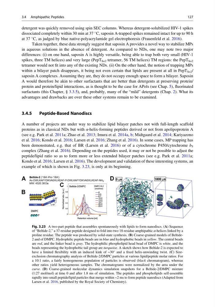
example of which is shown in Fig. 3.23, is only at its beginning.
Fig. 3.23 A two-part peptide that assembles spontaneously with lipids to form nanodiscs. (A) Sequence
of “Beltide-2,” a 37-residue peptide designed to fold into two 18-residue amphipathic α-helices linked by a
proline residue. The peptide was produced by solid-state synthesis. (B) Coarse-grained models of Beltide2 and of DMPC. Hydrophilic peptide beads are in blue and hydrophobic beads in yellow. The central beads
are red, and the linker bead is gray. The hydrophilic phospholipid head bead of DMPC is white, and the
beads representing the hydrophobic tail group are turquoise. A sketch shows how Beltide-2 is expected to
have a limited flexibility with an induced kink of ~30
and a fixed helix-unwinding twist. (C) Sizeexclusion chromatography analysis of Beltide-2/DMPC particles at various lipid/peptide molar ratios. For
a 10:1 ratio, a fairly homogeneous population of particles is observed (black chromatogram), whereas
other ratios yield heterogeneous samples. The chromatograms were normalized by the area under the
curve. (D) Coarse-grained molecular dynamics simulation snapshots for a Beltide-2/DMPC mixture
(1:27 mol/mol) at time 0 and after 1.8 ms of simulation. The peptides and phospholipids self-assemble
rapidly into small peptide/lipid particles that merge within ~2 ms to form peptide nanodiscs (Adapted from
Larsen et al. 2016, published by the Royal Society of Chemistry).
3.4 Amphipathic Peptides
127
dissociated completely within 30 min at 37
C, saposin A-trapped spikes remained intact for up to 90 h
at 37
C, as judged by blue native-polyacrylamide gel electrophoresis (Frauenfeld et al. 2016).
Taken together, these data strongly suggest that saposin A provides a novel way to stabilize MPs
in aqueous solutions in the absence of detergent. As compared to NDs, one may note two major
differences: (i) on one hand, saposin A is highly versatile, being able to trap both very small (HIV-1
spikes, three TM helices) and very large (PepT So2 tetramer, 56 TM helices) TM regions: the PepT So2
tetramer would not fit into any of the existing NDs. (ii) On the other hand, the notion of trapping MPs
within a bilayer patch disappears, it being not even certain that lipids are present at all in PepT So2 /
saposin A complexes. Assuming they are, they do not occupy enough space to form a bilayer. Saposin
A would therefore be akin to other surfactants that are better than detergents at preserving protein/
protein and protein/lipid interactions, as is thought to be the case for APols (see Chap. 5), fluorinated
surfactants (this Chapter, § 3.3.5), and, probably, many of the “mild” detergents (Chap. 2). What its
advantages and drawbacks are over these other systems remain to be examined.
3.4.5
Peptide-Based Nanodiscs
A number of projects are under way to stabilize lipid bilayer patches not with full-length scaffold
proteins as in classical NDs but with α-helix-forming peptides derived or not from apolipoprotein A
(see e.g. Park et al. 2011a; Zhao et al. 2013; Imura et al. 2014a, b; Midtgaard et al. 2014; Kariyazono
et al. 2016; Kondo et al. 2016; Larsen et al. 2016; Zhang et al. 2016). In some cases, MP trapping has
been demonstrated, e.g. that of BR (Larsen et al. 2016) or of a cytochrome P450/cytochrome b 5
complex (Zhang et al. 2016). Depending on the peptides used, it may or not be possible to adjust the
peptide/lipid ratio so as to form more or less extended bilayer patches (see e.g. Park et al. 2011a;
Kondo et al. 2016; Larsen et al. 2016). The development and validation of these interesting systems, an
example of which is shown in Fig. 3.23, is only at its beginning.
Fig. 3.23 A two-part peptide that assembles spontaneously with lipids to form nanodiscs. (A) Sequence
of “Beltide-2,” a 37-residue peptide designed to fold into two 18-residue amphipathic α-helices linked by a
proline residue. The peptide was produced by solid-state synthesis. (B) Coarse-grained models of Beltide2 and of DMPC. Hydrophilic peptide beads are in blue and hydrophobic beads in yellow. The central beads
are red, and the linker bead is gray. The hydrophilic phospholipid head bead of DMPC is white, and the
beads representing the hydrophobic tail group are turquoise. A sketch shows how Beltide-2 is expected to
have a limited flexibility with an induced kink of ~30
and a fixed helix-unwinding twist. (C) Sizeexclusion chromatography analysis of Beltide-2/DMPC particles at various lipid/peptide molar ratios. For
a 10:1 ratio, a fairly homogeneous population of particles is observed (black chromatogram), whereas
other ratios yield heterogeneous samples. The chromatograms were normalized by the area under the
curve. (D) Coarse-grained molecular dynamics simulation snapshots for a Beltide-2/DMPC mixture
(1:27 mol/mol) at time 0 and after 1.8 ms of simulation. The peptides and phospholipids self-assemble
rapidly into small peptide/lipid particles that merge within ~2 ms to form peptide nanodiscs (Adapted from
Larsen et al. 2016, published by the Royal Society of Chemistry).
3.4 Amphipathic Peptides
127