

gp120 trimer and an integral gp41 one, each gp41 monomer featuring one TM α-helix (Frauenfeld
et al. 2016; in this work, saposin A/lipid complexes are called Salipro® – short for saposin-lipoprotein
and a brand name – nanoparticles). Detergent-purified MPs were supplemented with saposin A in the
presence (in the case of T2 and PepT So2 ) or absence (HIV virus-like particles) of lipids, after which the
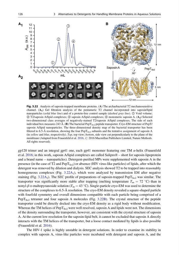
detergent was removed by dilution and dialysis. SEC analysis showed T2 to be trapped into reasonably
homogeneous complexes (Fig. 3.22A 1 ), which were analyzed by transmission EM after negative
staining (Fig. 3.22A 2 ). The SEC profile of preparations of saposin-trapped PepT So2 was similar. The
transporter was significantly more stable after trapping (melting temperature T m ¼ 72
C) than in
nonyl-β-D-maltopyranoside solution (T m ¼ 43
C). Single-particle cryo-EM was used to determine the
structure of the complexes to 6.5-Å resolution. The cryo-EM density revealed a square-shaped particle
with fourfold symmetry and overall dimensions compatible with each particle being comprised of a
PepT So2 tetramer and four saposin A molecules (Fig. 3.22B). The crystal structure of the peptide
transporter could be directly docked into the cryo-EM density as a rigid body without modification.
Whereas the TM helices of PepT So2 were well resolved, saposin A and lipids were not. The dimensions
of the density surrounding the transporter, however, are consistent with the crystal structure of saposin
A. At the current low resolution for the saposin-lipid belt, it cannot be excluded that saposin A directly
interacts with the TM helices of the transporter, but a loose contact mediated by lipids is also possible
(Frauenfeld et al. 2016).
The HIV-1 spike is highly unstable in detergent solutions. In order to examine its stability in
complex with saposin A, virus-like particles were incubated with detergent and saposin A, and the
Fig. 3.22 Analysis of saposin-trapped membrane proteins. (A) The archaebacterial T2 mechanosensitive
channel. (A 1 ) Gel filtration analysis of the pentameric T2 channel incorporated into saposin/lipid
nanoparticles (solid blue line) and of a protein-free control sample (dashed gray line). ① Void volume;
② T2/saposin A/lipid complexes; ③ saposin A/lipid complexes; ④ monomeric saposin A. (A 2 ) Selected
two-dimensional class averages of negatively-stained T2/saposin A/lipid complexes. The side of each
individual box measures 243 Å. (B) The bacterial PepT So2 peptide transporter. Cryo-EM structure of PepT/
saposin A/lipid nanoparticles. The three-dimensional density map of the bacterial transporter has been
filtered to 6.5-Å resolution, showing the four PepT So2 subunits and the tentative assignment of saposin A
(in yellow and blue, respectively). Top, top view; bottom, side view cut perpendicularly to the plane of the
membrane (Adapted from Frauenfeld et al. 2016, # 2016 Macmillan Publishers Limited, Nature Methods.
All rights reserved).
126
3 Alternatives to Detergents for Handling Membrane Proteins in Aqueous Solutions
et al. 2016; in this work, saposin A/lipid complexes are called Salipro® – short for saposin-lipoprotein
and a brand name – nanoparticles). Detergent-purified MPs were supplemented with saposin A in the
presence (in the case of T2 and PepT So2 ) or absence (HIV virus-like particles) of lipids, after which the
detergent was removed by dilution and dialysis. SEC analysis showed T2 to be trapped into reasonably
homogeneous complexes (Fig. 3.22A 1 ), which were analyzed by transmission EM after negative
staining (Fig. 3.22A 2 ). The SEC profile of preparations of saposin-trapped PepT So2 was similar. The
transporter was significantly more stable after trapping (melting temperature T m ¼ 72
C) than in
nonyl-β-D-maltopyranoside solution (T m ¼ 43
C). Single-particle cryo-EM was used to determine the
structure of the complexes to 6.5-Å resolution. The cryo-EM density revealed a square-shaped particle
with fourfold symmetry and overall dimensions compatible with each particle being comprised of a
PepT So2 tetramer and four saposin A molecules (Fig. 3.22B). The crystal structure of the peptide
transporter could be directly docked into the cryo-EM density as a rigid body without modification.
Whereas the TM helices of PepT So2 were well resolved, saposin A and lipids were not. The dimensions
of the density surrounding the transporter, however, are consistent with the crystal structure of saposin
A. At the current low resolution for the saposin-lipid belt, it cannot be excluded that saposin A directly
interacts with the TM helices of the transporter, but a loose contact mediated by lipids is also possible
(Frauenfeld et al. 2016).
The HIV-1 spike is highly unstable in detergent solutions. In order to examine its stability in
complex with saposin A, virus-like particles were incubated with detergent and saposin A, and the
Fig. 3.22 Analysis of saposin-trapped membrane proteins. (A) The archaebacterial T2 mechanosensitive
channel. (A 1 ) Gel filtration analysis of the pentameric T2 channel incorporated into saposin/lipid
nanoparticles (solid blue line) and of a protein-free control sample (dashed gray line). ① Void volume;
② T2/saposin A/lipid complexes; ③ saposin A/lipid complexes; ④ monomeric saposin A. (A 2 ) Selected
two-dimensional class averages of negatively-stained T2/saposin A/lipid complexes. The side of each
individual box measures 243 Å. (B) The bacterial PepT So2 peptide transporter. Cryo-EM structure of PepT/
saposin A/lipid nanoparticles. The three-dimensional density map of the bacterial transporter has been
filtered to 6.5-Å resolution, showing the four PepT So2 subunits and the tentative assignment of saposin A
(in yellow and blue, respectively). Top, top view; bottom, side view cut perpendicularly to the plane of the
membrane (Adapted from Frauenfeld et al. 2016, # 2016 Macmillan Publishers Limited, Nature Methods.
All rights reserved).
126
3 Alternatives to Detergents for Handling Membrane Proteins in Aqueous Solutions