

A/LDAO complexes (Fig. 3.20B 1 ). As with LDAO, the lipid tails are held within the central cavity of
the complex, where they interact with the hydrophobic faces of the saposin A protomers (Fig. 3.20B 2 ).
A representative snapshot reveals that the lipids are arranged in two leaflets as a mini-bilayer
(Fig. 3.20B 3 ), with more lipid molecules tending to lie in one leaflet than in the other. As noted by
the authors of the study, the arrangement observed in the crystal structure may well represent only one
of the configurations that saposin A/detergent or saposin A/lipid complexes may adopt in solution
(Popovic et al. 2012). This hypothesis has been vindicated by more detailed experimental studies
(Li et al. 2016b; see below). The so-called Sap A discs or picodiscs formed by saposin A-solubilized
lipids have been exploited as a convenient formulation for presenting glycolipids to hydrolases or
lipid-binding proteins (Leney et al. 2015; Li et al. 2016a).
Further studies, using catch-and-release electrospray ionization mass spectrometry (CaR-ESI-MS),
size-exclusion chromatography/multi-angle laser light scattering (SEC-MALLS), and MD, do show that
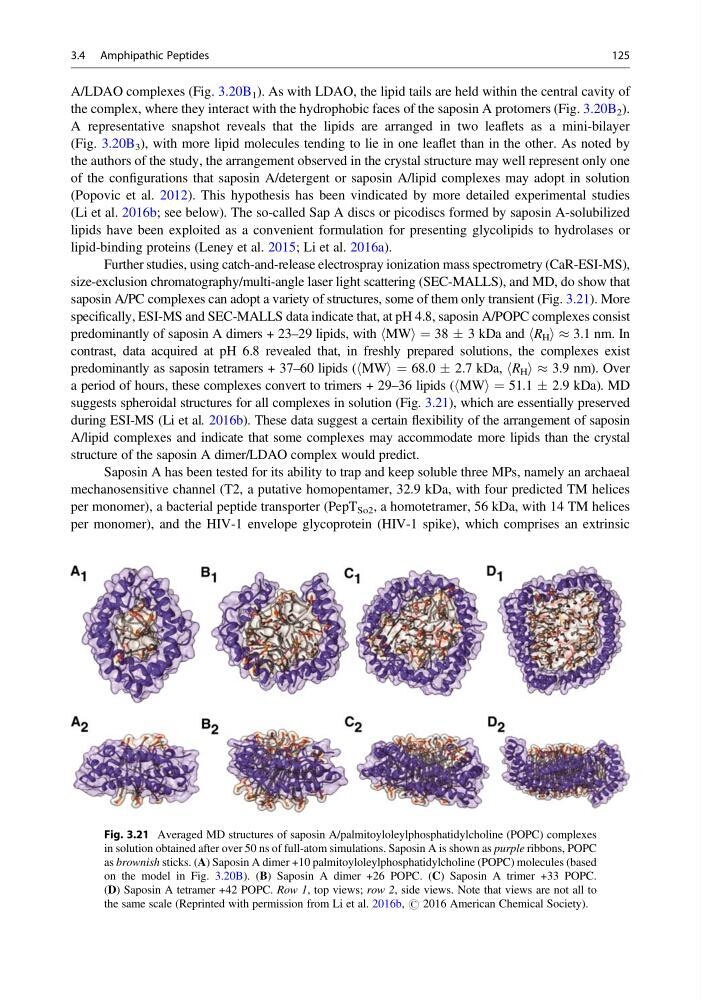
saposin A/PC complexes can adopt a variety of structures, some of them only transient (Fig. 3.21). More
specifically, ESI-MS and SEC-MALLS data indicate that, at pH 4.8, saposin A/POPC complexes consist
predominantly of saposin A dimers + 23–29 lipids, with hMWi ¼ 38 Æ 3 kDa and hR H i % 3.1 nm. In
contrast, data acquired at pH 6.8 revealed that, in freshly prepared solutions, the complexes exist
predominantly as saposin tetramers + 37–60 lipids (hMWi ¼ 68.0 Æ 2.7 kDa, hR H i % 3.9 nm). Over
a period of hours, these complexes convert to trimers + 29–36 lipids (hMWi ¼ 51.1 Æ 2.9 kDa). MD
suggests spheroidal structures for all complexes in solution (Fig. 3.21), which are essentially preserved
during ESI-MS (Li et al. 2016b). These data suggest a certain flexibility of the arrangement of saposin
A/lipid complexes and indicate that some complexes may accommodate more lipids than the crystal
structure of the saposin A dimer/LDAO complex would predict.
Saposin A has been tested for its ability to trap and keep soluble three MPs, namely an archaeal
mechanosensitive channel (T2, a putative homopentamer, 32.9 kDa, with four predicted TM helices
per monomer), a bacterial peptide transporter (PepT So2 , a homotetramer, 56 kDa, with 14 TM helices
per monomer), and the HIV-1 envelope glycoprotein (HIV-1 spike), which comprises an extrinsic
Fig. 3.21 Averaged MD structures of saposin A/palmitoyloleylphosphatidylcholine (POPC) complexes
in solution obtained after over 50 ns of full-atom simulations. Saposin A is shown as purple ribbons, POPC
as brownish sticks. (A) Saposin A dimer +10 palmitoyloleylphosphatidylcholine (POPC) molecules (based
on the model in Fig. 3.20B). (B) Saposin A dimer +26 POPC. (C) Saposin A trimer +33 POPC.
(D) Saposin A tetramer +42 POPC. Row 1, top views; row 2, side views. Note that views are not all to
the same scale (Reprinted with permission from Li et al. 2016b, # 2016 American Chemical Society).
3.4 Amphipathic Peptides
125
the complex, where they interact with the hydrophobic faces of the saposin A protomers (Fig. 3.20B 2 ).
A representative snapshot reveals that the lipids are arranged in two leaflets as a mini-bilayer
(Fig. 3.20B 3 ), with more lipid molecules tending to lie in one leaflet than in the other. As noted by
the authors of the study, the arrangement observed in the crystal structure may well represent only one
of the configurations that saposin A/detergent or saposin A/lipid complexes may adopt in solution
(Popovic et al. 2012). This hypothesis has been vindicated by more detailed experimental studies
(Li et al. 2016b; see below). The so-called Sap A discs or picodiscs formed by saposin A-solubilized
lipids have been exploited as a convenient formulation for presenting glycolipids to hydrolases or
lipid-binding proteins (Leney et al. 2015; Li et al. 2016a).
Further studies, using catch-and-release electrospray ionization mass spectrometry (CaR-ESI-MS),
size-exclusion chromatography/multi-angle laser light scattering (SEC-MALLS), and MD, do show that
saposin A/PC complexes can adopt a variety of structures, some of them only transient (Fig. 3.21). More
specifically, ESI-MS and SEC-MALLS data indicate that, at pH 4.8, saposin A/POPC complexes consist
predominantly of saposin A dimers + 23–29 lipids, with hMWi ¼ 38 Æ 3 kDa and hR H i % 3.1 nm. In
contrast, data acquired at pH 6.8 revealed that, in freshly prepared solutions, the complexes exist
predominantly as saposin tetramers + 37–60 lipids (hMWi ¼ 68.0 Æ 2.7 kDa, hR H i % 3.9 nm). Over
a period of hours, these complexes convert to trimers + 29–36 lipids (hMWi ¼ 51.1 Æ 2.9 kDa). MD
suggests spheroidal structures for all complexes in solution (Fig. 3.21), which are essentially preserved
during ESI-MS (Li et al. 2016b). These data suggest a certain flexibility of the arrangement of saposin
A/lipid complexes and indicate that some complexes may accommodate more lipids than the crystal
structure of the saposin A dimer/LDAO complex would predict.
Saposin A has been tested for its ability to trap and keep soluble three MPs, namely an archaeal
mechanosensitive channel (T2, a putative homopentamer, 32.9 kDa, with four predicted TM helices
per monomer), a bacterial peptide transporter (PepT So2 , a homotetramer, 56 kDa, with 14 TM helices
per monomer), and the HIV-1 envelope glycoprotein (HIV-1 spike), which comprises an extrinsic
Fig. 3.21 Averaged MD structures of saposin A/palmitoyloleylphosphatidylcholine (POPC) complexes
in solution obtained after over 50 ns of full-atom simulations. Saposin A is shown as purple ribbons, POPC
as brownish sticks. (A) Saposin A dimer +10 palmitoyloleylphosphatidylcholine (POPC) molecules (based
on the model in Fig. 3.20B). (B) Saposin A dimer +26 POPC. (C) Saposin A trimer +33 POPC.
(D) Saposin A tetramer +42 POPC. Row 1, top views; row 2, side views. Note that views are not all to
the same scale (Reprinted with permission from Li et al. 2016b, # 2016 American Chemical Society).
3.4 Amphipathic Peptides
125