

Indeed, following incubation of liposomes of various compositions with saposin A, SEC analysis
reveals the presence of particles in the 35 to 45-kDa range, with hydrodynamic radii R H % 3.2 nm
(Popovic et al. 2012). The particles obtained by incubating saposin A with liposomes made from egg
phosphatidylcholine (PC) contain 5 Æ 1 lipids per saposin A chain, leading to the suggestion that they
comprise two copies of saposin A and 8–12 lipids (Popovic et al. 2012).
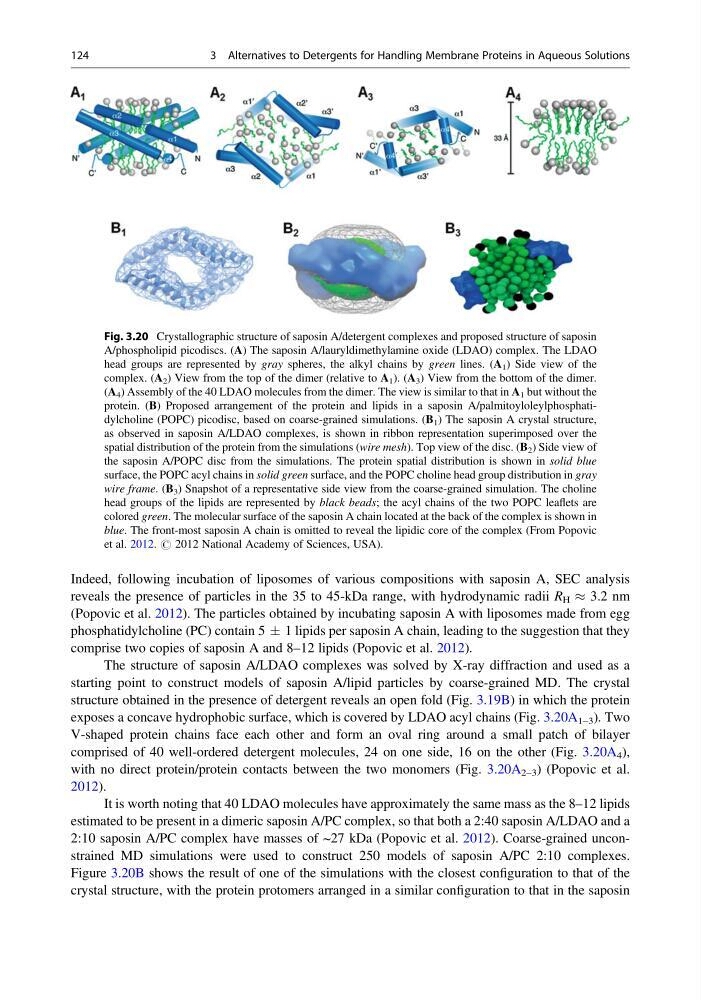
The structure of saposin A/LDAO complexes was solved by X-ray diffraction and used as a
starting point to construct models of saposin A/lipid particles by coarse-grained MD. The crystal
structure obtained in the presence of detergent reveals an open fold (Fig. 3.19B) in which the protein
exposes a concave hydrophobic surface, which is covered by LDAO acyl chains (Fig. 3.20A 1–3 ). Two
V-shaped protein chains face each other and form an oval ring around a small patch of bilayer
comprised of 40 well-ordered detergent molecules, 24 on one side, 16 on the other (Fig. 3.20A 4 ),
with no direct protein/protein contacts between the two monomers (Fig. 3.20A 2–3 ) (Popovic et al.
2012).
It is worth noting that 40 LDAO molecules have approximately the same mass as the 8–12 lipids
estimated to be present in a dimeric saposin A/PC complex, so that both a 2:40 saposin A/LDAO and a
2:10 saposin A/PC complex have masses of ~27 kDa (Popovic et al. 2012). Coarse-grained unconstrained MD simulations were used to construct 250 models of saposin A/PC 2:10 complexes.
Figure 3.20B shows the result of one of the simulations with the closest configuration to that of the
crystal structure, with the protein protomers arranged in a similar configuration to that in the saposin
Fig. 3.20 Crystallographic structure of saposin A/detergent complexes and proposed structure of saposin
A/phospholipid picodiscs. (A) The saposin A/lauryldimethylamine oxide (LDAO) complex. The LDAO
head groups are represented by gray spheres, the alkyl chains by green lines. (A 1 ) Side view of the
complex. (A 2 ) View from the top of the dimer (relative to A 1 ). (A 3 ) View from the bottom of the dimer.
(A 4 ) Assembly of the 40 LDAO molecules from the dimer. The view is similar to that in A 1 but without the
protein. (B) Proposed arrangement of the protein and lipids in a saposin A/palmitoyloleylphosphatidylcholine (POPC) picodisc, based on coarse-grained simulations. (B 1 ) The saposin A crystal structure,
as observed in saposin A/LDAO complexes, is shown in ribbon representation superimposed over the
spatial distribution of the protein from the simulations (wire mesh). Top view of the disc. (B 2 ) Side view of
the saposin A/POPC disc from the simulations. The protein spatial distribution is shown in solid blue
surface, the POPC acyl chains in solid green surface, and the POPC choline head group distribution in gray
wire frame. (B 3 ) Snapshot of a representative side view from the coarse-grained simulation. The choline
head groups of the lipids are represented by black beads; the acyl chains of the two POPC leaflets are
colored green. The molecular surface of the saposin A chain located at the back of the complex is shown in
blue. The front-most saposin A chain is omitted to reveal the lipidic core of the complex (From Popovic
et al. 2012. # 2012 National Academy of Sciences, USA).
124
3 Alternatives to Detergents for Handling Membrane Proteins in Aqueous Solutions
reveals the presence of particles in the 35 to 45-kDa range, with hydrodynamic radii R H % 3.2 nm
(Popovic et al. 2012). The particles obtained by incubating saposin A with liposomes made from egg
phosphatidylcholine (PC) contain 5 Æ 1 lipids per saposin A chain, leading to the suggestion that they
comprise two copies of saposin A and 8–12 lipids (Popovic et al. 2012).
The structure of saposin A/LDAO complexes was solved by X-ray diffraction and used as a
starting point to construct models of saposin A/lipid particles by coarse-grained MD. The crystal
structure obtained in the presence of detergent reveals an open fold (Fig. 3.19B) in which the protein
exposes a concave hydrophobic surface, which is covered by LDAO acyl chains (Fig. 3.20A 1–3 ). Two
V-shaped protein chains face each other and form an oval ring around a small patch of bilayer
comprised of 40 well-ordered detergent molecules, 24 on one side, 16 on the other (Fig. 3.20A 4 ),
with no direct protein/protein contacts between the two monomers (Fig. 3.20A 2–3 ) (Popovic et al.
2012).
It is worth noting that 40 LDAO molecules have approximately the same mass as the 8–12 lipids
estimated to be present in a dimeric saposin A/PC complex, so that both a 2:40 saposin A/LDAO and a
2:10 saposin A/PC complex have masses of ~27 kDa (Popovic et al. 2012). Coarse-grained unconstrained MD simulations were used to construct 250 models of saposin A/PC 2:10 complexes.
Figure 3.20B shows the result of one of the simulations with the closest configuration to that of the
crystal structure, with the protein protomers arranged in a similar configuration to that in the saposin
Fig. 3.20 Crystallographic structure of saposin A/detergent complexes and proposed structure of saposin
A/phospholipid picodiscs. (A) The saposin A/lauryldimethylamine oxide (LDAO) complex. The LDAO
head groups are represented by gray spheres, the alkyl chains by green lines. (A 1 ) Side view of the
complex. (A 2 ) View from the top of the dimer (relative to A 1 ). (A 3 ) View from the bottom of the dimer.
(A 4 ) Assembly of the 40 LDAO molecules from the dimer. The view is similar to that in A 1 but without the
protein. (B) Proposed arrangement of the protein and lipids in a saposin A/palmitoyloleylphosphatidylcholine (POPC) picodisc, based on coarse-grained simulations. (B 1 ) The saposin A crystal structure,
as observed in saposin A/LDAO complexes, is shown in ribbon representation superimposed over the
spatial distribution of the protein from the simulations (wire mesh). Top view of the disc. (B 2 ) Side view of
the saposin A/POPC disc from the simulations. The protein spatial distribution is shown in solid blue
surface, the POPC acyl chains in solid green surface, and the POPC choline head group distribution in gray
wire frame. (B 3 ) Snapshot of a representative side view from the coarse-grained simulation. The choline
head groups of the lipids are represented by black beads; the acyl chains of the two POPC leaflets are
colored green. The molecular surface of the saposin A chain located at the back of the complex is shown in
blue. The front-most saposin A chain is omitted to reveal the lipidic core of the complex (From Popovic
et al. 2012. # 2012 National Academy of Sciences, USA).
124
3 Alternatives to Detergents for Handling Membrane Proteins in Aqueous Solutions