

peptide backbones and the higher rigidity of the peptides as compared to fatty acyl chains (von
Maltzahn et al. 2003). When using DPSs to trap MPs, an important issue is therefore whether one is
dealing with large aggregates or small complexes. Photosystem I reaction centers transferred from
detergent solution to DPSs were found to be associated with large or small vesicles, depending on the
peptide used (Matsumoto et al. 2009). The GPCR rhodopsin (Rho) was stabilized upon transfer from
either DDM or OG to either detergent/DPS mixtures or pure DPSs, whether in the presence or absence
of lipids (Zhao et al. 2006). Stabilization was markedly higher in the presence of lipids, strongly
suggesting that Rho/lipid interactions persist in the presence of DPSs. The size of the objects formed
was not reported.
DPSs can form micelles (Koutsopoulos et al. 2012), which would lead one to believe that small
MP/DPS complexes can be obtained, but none seems to have been described yet, leaving it uncertain
whether MP/DPS complexes can be used for NMR studies or crystallization attempts.
3.4.4
Stabilizing Membrane Proteins by Complexation with Saposin A
(“Picodiscs,” “Sap A discs,” “Salipro® Nanoparticles”)
Saposins are small (~80 residues), nonenzymatic proteins required for the breakdown of glycosphingolipids within the lysosome (Kolter and Sandhoff 2005). Each saposin activates the breakdown of
particular lipid substrates by facilitating the access of the lipid head groups to the active site of
hydrolases. Saposins are thought to act by solubilizing the lipid substrates or simply by destabilizing
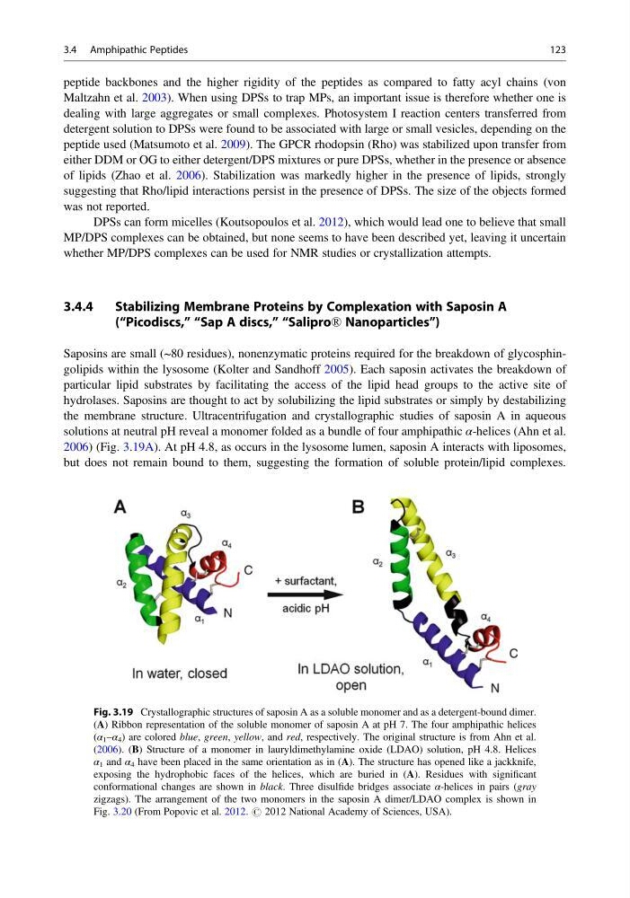
the membrane structure. Ultracentrifugation and crystallographic studies of saposin A in aqueous
solutions at neutral pH reveal a monomer folded as a bundle of four amphipathic α-helices (Ahn et al.
2006) (Fig. 3.19A). At pH 4.8, as occurs in the lysosome lumen, saposin A interacts with liposomes,
but does not remain bound to them, suggesting the formation of soluble protein/lipid complexes.
Fig. 3.19 Crystallographic structures of saposin A as a soluble monomer and as a detergent-bound dimer.
(A) Ribbon representation of the soluble monomer of saposin A at pH 7. The four amphipathic helices
(α 1 –α 4 ) are colored blue, green, yellow, and red, respectively. The original structure is from Ahn et al.
(2006). (B) Structure of a monomer in lauryldimethylamine oxide (LDAO) solution, pH 4.8. Helices
α 1 and α 4 have been placed in the same orientation as in (A). The structure has opened like a jackknife,
exposing the hydrophobic faces of the helices, which are buried in (A). Residues with significant
conformational changes are shown in black. Three disulfide bridges associate α-helices in pairs (gray
zigzags). The arrangement of the two monomers in the saposin A dimer/LDAO complex is shown in
Fig. 3.20 (From Popovic et al. 2012. # 2012 National Academy of Sciences, USA).
3.4 Amphipathic Peptides
123
Maltzahn et al. 2003). When using DPSs to trap MPs, an important issue is therefore whether one is
dealing with large aggregates or small complexes. Photosystem I reaction centers transferred from
detergent solution to DPSs were found to be associated with large or small vesicles, depending on the
peptide used (Matsumoto et al. 2009). The GPCR rhodopsin (Rho) was stabilized upon transfer from
either DDM or OG to either detergent/DPS mixtures or pure DPSs, whether in the presence or absence
of lipids (Zhao et al. 2006). Stabilization was markedly higher in the presence of lipids, strongly
suggesting that Rho/lipid interactions persist in the presence of DPSs. The size of the objects formed
was not reported.
DPSs can form micelles (Koutsopoulos et al. 2012), which would lead one to believe that small
MP/DPS complexes can be obtained, but none seems to have been described yet, leaving it uncertain
whether MP/DPS complexes can be used for NMR studies or crystallization attempts.
3.4.4
Stabilizing Membrane Proteins by Complexation with Saposin A
(“Picodiscs,” “Sap A discs,” “Salipro® Nanoparticles”)
Saposins are small (~80 residues), nonenzymatic proteins required for the breakdown of glycosphingolipids within the lysosome (Kolter and Sandhoff 2005). Each saposin activates the breakdown of
particular lipid substrates by facilitating the access of the lipid head groups to the active site of
hydrolases. Saposins are thought to act by solubilizing the lipid substrates or simply by destabilizing
the membrane structure. Ultracentrifugation and crystallographic studies of saposin A in aqueous
solutions at neutral pH reveal a monomer folded as a bundle of four amphipathic α-helices (Ahn et al.
2006) (Fig. 3.19A). At pH 4.8, as occurs in the lysosome lumen, saposin A interacts with liposomes,
but does not remain bound to them, suggesting the formation of soluble protein/lipid complexes.
Fig. 3.19 Crystallographic structures of saposin A as a soluble monomer and as a detergent-bound dimer.
(A) Ribbon representation of the soluble monomer of saposin A at pH 7. The four amphipathic helices
(α 1 –α 4 ) are colored blue, green, yellow, and red, respectively. The original structure is from Ahn et al.
(2006). (B) Structure of a monomer in lauryldimethylamine oxide (LDAO) solution, pH 4.8. Helices
α 1 and α 4 have been placed in the same orientation as in (A). The structure has opened like a jackknife,
exposing the hydrophobic faces of the helices, which are buried in (A). Residues with significant
conformational changes are shown in black. Three disulfide bridges associate α-helices in pairs (gray
zigzags). The arrangement of the two monomers in the saposin A dimer/LDAO complex is shown in
Fig. 3.20 (From Popovic et al. 2012. # 2012 National Academy of Sciences, USA).
3.4 Amphipathic Peptides
123