

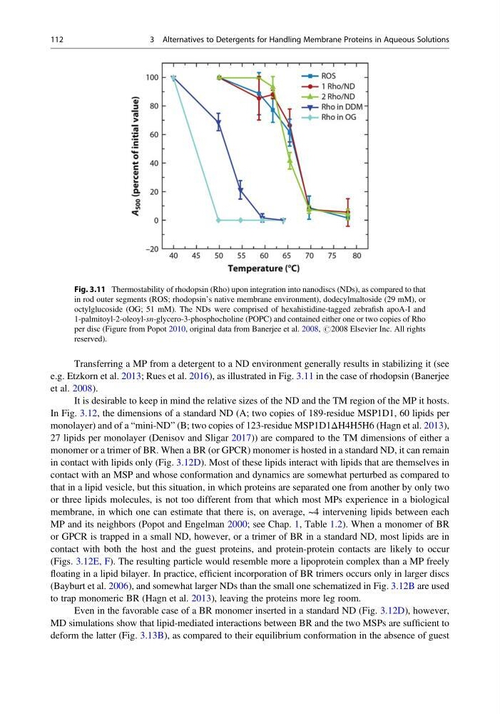
Transferring a MP from a detergent to a ND environment generally results in stabilizing it (see
e.g. Etzkorn et al. 2013; Rues et al. 2016), as illustrated in Fig. 3.11 in the case of rhodopsin (Banerjee
et al. 2008).
It is desirable to keep in mind the relative sizes of the ND and the TM region of the MP it hosts.
In Fig. 3.12, the dimensions of a standard ND (A; two copies of 189-residue MSP1D1, 60 lipids per
monolayer) and of a “mini-ND” (B; two copies of 123-residue MSP1D1ΔH4H5H6 (Hagn et al. 2013),
27 lipids per monolayer (Denisov and Sligar 2017)) are compared to the TM dimensions of either a
monomer or a trimer of BR. When a BR (or GPCR) monomer is hosted in a standard ND, it can remain
in contact with lipids only (Fig. 3.12D). Most of these lipids interact with lipids that are themselves in
contact with an MSP and whose conformation and dynamics are somewhat perturbed as compared to
that in a lipid vesicle, but this situation, in which proteins are separated one from another by only two
or three lipids molecules, is not too different from that which most MPs experience in a biological
membrane, in which one can estimate that there is, on average, ~4 intervening lipids between each
MP and its neighbors (Popot and Engelman 2000; see Chap. 1, Table 1.2). When a monomer of BR
or GPCR is trapped in a small ND, however, or a trimer of BR in a standard ND, most lipids are in
contact with both the host and the guest proteins, and protein-protein contacts are likely to occur
(Figs. 3.12E, F). The resulting particle would resemble more a lipoprotein complex than a MP freely
floating in a lipid bilayer. In practice, efficient incorporation of BR trimers occurs only in larger discs
(Bayburt et al. 2006), and somewhat larger NDs than the small one schematized in Fig. 3.12B are used
to trap monomeric BR (Hagn et al. 2013), leaving the proteins more leg room.
Even in the favorable case of a BR monomer inserted in a standard ND (Fig. 3.12D), however,
MD simulations show that lipid-mediated interactions between BR and the two MSPs are sufficient to
deform the latter (Fig. 3.13B), as compared to their equilibrium conformation in the absence of guest
Fig. 3.11 Thermostability of rhodopsin (Rho) upon integration into nanodiscs (NDs), as compared to that
in rod outer segments (ROS; rhodopsin’s native membrane environment), dodecylmaltoside (29 mM), or
octylglucoside (OG; 51 mM). The NDs were comprised of hexahistidine-tagged zebrafish apoA-I and
1-palmitoyl-2-oleoyl-sn-glycero-3-phosphocholine (POPC) and contained either one or two copies of Rho
per disc (Figure from Popot 2010, original data from Banerjee et al. 2008, #2008 Elsevier Inc. All rights
reserved).
112
3 Alternatives to Detergents for Handling Membrane Proteins in Aqueous Solutions
e.g. Etzkorn et al. 2013; Rues et al. 2016), as illustrated in Fig. 3.11 in the case of rhodopsin (Banerjee
et al. 2008).
It is desirable to keep in mind the relative sizes of the ND and the TM region of the MP it hosts.
In Fig. 3.12, the dimensions of a standard ND (A; two copies of 189-residue MSP1D1, 60 lipids per
monolayer) and of a “mini-ND” (B; two copies of 123-residue MSP1D1ΔH4H5H6 (Hagn et al. 2013),
27 lipids per monolayer (Denisov and Sligar 2017)) are compared to the TM dimensions of either a
monomer or a trimer of BR. When a BR (or GPCR) monomer is hosted in a standard ND, it can remain
in contact with lipids only (Fig. 3.12D). Most of these lipids interact with lipids that are themselves in
contact with an MSP and whose conformation and dynamics are somewhat perturbed as compared to
that in a lipid vesicle, but this situation, in which proteins are separated one from another by only two
or three lipids molecules, is not too different from that which most MPs experience in a biological
membrane, in which one can estimate that there is, on average, ~4 intervening lipids between each
MP and its neighbors (Popot and Engelman 2000; see Chap. 1, Table 1.2). When a monomer of BR
or GPCR is trapped in a small ND, however, or a trimer of BR in a standard ND, most lipids are in
contact with both the host and the guest proteins, and protein-protein contacts are likely to occur
(Figs. 3.12E, F). The resulting particle would resemble more a lipoprotein complex than a MP freely
floating in a lipid bilayer. In practice, efficient incorporation of BR trimers occurs only in larger discs
(Bayburt et al. 2006), and somewhat larger NDs than the small one schematized in Fig. 3.12B are used
to trap monomeric BR (Hagn et al. 2013), leaving the proteins more leg room.
Even in the favorable case of a BR monomer inserted in a standard ND (Fig. 3.12D), however,
MD simulations show that lipid-mediated interactions between BR and the two MSPs are sufficient to
deform the latter (Fig. 3.13B), as compared to their equilibrium conformation in the absence of guest
Fig. 3.11 Thermostability of rhodopsin (Rho) upon integration into nanodiscs (NDs), as compared to that
in rod outer segments (ROS; rhodopsin’s native membrane environment), dodecylmaltoside (29 mM), or
octylglucoside (OG; 51 mM). The NDs were comprised of hexahistidine-tagged zebrafish apoA-I and
1-palmitoyl-2-oleoyl-sn-glycero-3-phosphocholine (POPC) and contained either one or two copies of Rho
per disc (Figure from Popot 2010, original data from Banerjee et al. 2008, #2008 Elsevier Inc. All rights
reserved).
112
3 Alternatives to Detergents for Handling Membrane Proteins in Aqueous Solutions