

determine the angle that the axis of an α-helix or β-barrel makes with the plane of the bicelle and to
place constraints on the angle that the axes of two helices make with one another. Magnetic alignment
also provides an access to dipolar coupling and chemical shift anisotropy measurements, which can be
directly employed for protein structural determination (see e.g. Ketchem et al. 1993, where alignment
was achieved by the glass plate method).
The phase diagram of bicelle-forming surfactant mixtures is complex, because the structure and
properties of the assemblies formed depend on the ratio between the small and large surfactants, on
their nature, on their overall concentration, on the temperature, the presence of salts, and so forth (see
e.g. Sternin et al. 2001; Harroun et al. 2005; Triba et al. 2005; van Dam et al. 2006; Ujwal and Bowie
2011; Warschawski et al. 2011; Dürr et al. 2012, 2013; Liebau et al. 2016). A phase diagram of DMPC/
diC 6 PC mixtures as a function of temperature and the mass ratio q of DMPC to diC 6 PC, established by
phosphorus NMR, is shown in Fig. 3.3, left, and some of the structures adopted schematically depicted
in Fig. 3.3, right. Bicelles in which the two lipids are fully segregated (B) occupy only part of the
diagram (below the melting temperature T m of DMPC, where its miscibility with diC 6 PC is minimal).
The other structures found include mixed micelles (M m ), at very low q; mixed bicelles (B m ), in the
bilayer region of which the two lipids partially mix; perforated vesicles or sheets (V p ); and
non-perforated vesicles (V m ) in which the two lipids are totally mixed. Raising the temperature of a
given mixture (red arrow in Fig. 3.3, left) increases the miscibility of the two lipids, which takes the
preparation through the various types of structures. Care should therefore be brought to choosing
experimental conditions so as to remain in that region of the mixture’s phase diagram where bicelles of
the desired size will form, e.g. the small so-called “isotropic” bicelles – meaning “tumbling
isotropically” – used for solution NMR (B in Fig. 3.3) or the large mixed bicelles (B m in Fig. 3.3)
that align best in magnetic fields and are used for solid-state NMR applications. Excessive dilution
should be avoided, for instance, or the bicelles will turn into vesicles. This is a serious experimental
constraint, avoided by nanodiscs or amphipols. On the contrary, the transition from bicelles to
perforated or non-perforated vesicles or sheets probably plays a useful role when crystallizing MPs
from a bicellar preparation (see § 3.2.2). The way detergent solutions, isotropically tumbling and
aligned bicelles, and lipid vesicles complement each other in the study of the structure and dynamics of
MPs and their amenability to the various forms of NMR spectroscopy are schematically illustrated in
Fig. 3.4.
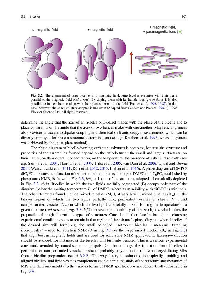
Fig. 3.2 The alignment of large bicelles in a magnetic field. Pure bicelles organize with their plane
parallel to the magnetic field (red arrow). By doping them with lanthanide ions (green dots), it is also
possible to induce them to align with their planes normal to the field (Prosser et al. 1996, 1998). In this
case, however, the exact structure adopted is uncertain (Adapted from Sanders and Prosser 1998. # 1998
Elsevier Science Ltd. All rights reserved).
3.2 Bicelles
101
place constraints on the angle that the axes of two helices make with one another. Magnetic alignment
also provides an access to dipolar coupling and chemical shift anisotropy measurements, which can be
directly employed for protein structural determination (see e.g. Ketchem et al. 1993, where alignment
was achieved by the glass plate method).
The phase diagram of bicelle-forming surfactant mixtures is complex, because the structure and
properties of the assemblies formed depend on the ratio between the small and large surfactants, on
their nature, on their overall concentration, on the temperature, the presence of salts, and so forth (see
e.g. Sternin et al. 2001; Harroun et al. 2005; Triba et al. 2005; van Dam et al. 2006; Ujwal and Bowie
2011; Warschawski et al. 2011; Dürr et al. 2012, 2013; Liebau et al. 2016). A phase diagram of DMPC/
diC 6 PC mixtures as a function of temperature and the mass ratio q of DMPC to diC 6 PC, established by
phosphorus NMR, is shown in Fig. 3.3, left, and some of the structures adopted schematically depicted
in Fig. 3.3, right. Bicelles in which the two lipids are fully segregated (B) occupy only part of the
diagram (below the melting temperature T m of DMPC, where its miscibility with diC 6 PC is minimal).
The other structures found include mixed micelles (M m ), at very low q; mixed bicelles (B m ), in the
bilayer region of which the two lipids partially mix; perforated vesicles or sheets (V p ); and
non-perforated vesicles (V m ) in which the two lipids are totally mixed. Raising the temperature of a
given mixture (red arrow in Fig. 3.3, left) increases the miscibility of the two lipids, which takes the
preparation through the various types of structures. Care should therefore be brought to choosing
experimental conditions so as to remain in that region of the mixture’s phase diagram where bicelles of
the desired size will form, e.g. the small so-called “isotropic” bicelles – meaning “tumbling
isotropically” – used for solution NMR (B in Fig. 3.3) or the large mixed bicelles (B m in Fig. 3.3)
that align best in magnetic fields and are used for solid-state NMR applications. Excessive dilution
should be avoided, for instance, or the bicelles will turn into vesicles. This is a serious experimental
constraint, avoided by nanodiscs or amphipols. On the contrary, the transition from bicelles to
perforated or non-perforated vesicles or sheets probably plays a useful role when crystallizing MPs
from a bicellar preparation (see § 3.2.2). The way detergent solutions, isotropically tumbling and
aligned bicelles, and lipid vesicles complement each other in the study of the structure and dynamics of
MPs and their amenability to the various forms of NMR spectroscopy are schematically illustrated in
Fig. 3.4.
Fig. 3.2 The alignment of large bicelles in a magnetic field. Pure bicelles organize with their plane
parallel to the magnetic field (red arrow). By doping them with lanthanide ions (green dots), it is also
possible to induce them to align with their planes normal to the field (Prosser et al. 1996, 1998). In this
case, however, the exact structure adopted is uncertain (Adapted from Sanders and Prosser 1998. # 1998
Elsevier Science Ltd. All rights reserved).
3.2 Bicelles
101