

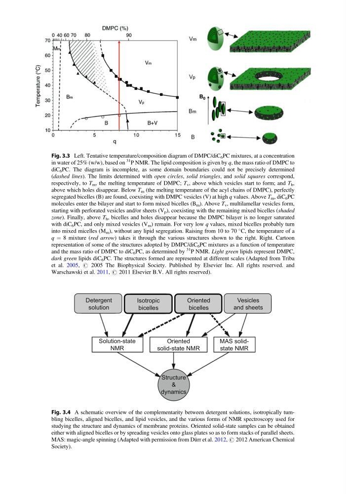
Fig. 3.3 Left. Tentative temperature/composition diagram of DMPC/diC 6 PC mixtures, at a concentration
in water of 25% (w/w), based on
31
P NMR. The lipid composition is given by q, the mass ratio of DMPC to
diC 6 PC. The diagram is incomplete, as some domain boundaries could not be precisely determined
(dashed lines). The limits determined with open circles, solid triangles, and solid squares correspond,
respectively, to T m , the melting temperature of DMPC; T v , above which vesicles start to form; and T h ,
above which holes disappear. Below T m (the melting temperature of the acyl chains of DMPC), perfectly
segregated bicelles (B) are found, coexisting with DMPC vesicles (V) at high q values. Above T m , diC 6 PC
molecules enter the bilayer and start to form mixed bicelles (B m ). Above T v , multilamellar vesicles form,
starting with perforated vesicles and/or sheets (V p ), coexisting with the remaining mixed bicelles (shaded
zone). Finally, above T h , bicelles and holes disappear because the DMPC bilayer is no longer saturated
with diC 6 PC, and only mixed vesicles (V m ) remain. For very low q values, mixed bicelles probably turn
into mixed micelles (M m ), without any lipid segregation. Raising from 10 to 70
C, the temperature of a
q ¼ 8 mixture (red arrow) takes it through the various structures shown to the right. Right. Cartoon
representation of some of the structures adopted by DMPC/diC 6 PC mixtures as a function of temperature
and the mass ratio of DMPC to diC 6 PC, as determined by
31
P NMR. Light green lipids represent DMPC,
dark green lipids diC 6 PC. The structures formed are represented at different scales (Adapted from Triba
et al. 2005, # 2005 The Biophysical Society. Published by Elsevier Inc. All rights reserved. and
Warschawski et al. 2011, # 2011 Elsevier B.V. All rights reserved).
Oriented
solid-state NMR
MAS solidstate NMR
Solution-state
NMR
Vesicles
and sheets
Detergent
solution
Oriented
bicelles
Isotropic
bicelles
Structure
&
dynamics
Fig. 3.4 A schematic overview of the complementarity between detergent solutions, isotropically tumbling bicelles, aligned bicelles, and lipid vesicles, and the various forms of NMR spectroscopy used for
studying the structure and dynamics of membrane proteins. Oriented solid-state samples can be obtained
either with aligned bicelles or by spreading vesicles onto glass plates so as to form stacks of parallel sheets.
MAS: magic-angle spinning (Adapted with permission from Dürr et al. 2012, # 2012 American Chemical
Society).
in water of 25% (w/w), based on
31
P NMR. The lipid composition is given by q, the mass ratio of DMPC to
diC 6 PC. The diagram is incomplete, as some domain boundaries could not be precisely determined
(dashed lines). The limits determined with open circles, solid triangles, and solid squares correspond,
respectively, to T m , the melting temperature of DMPC; T v , above which vesicles start to form; and T h ,
above which holes disappear. Below T m (the melting temperature of the acyl chains of DMPC), perfectly
segregated bicelles (B) are found, coexisting with DMPC vesicles (V) at high q values. Above T m , diC 6 PC
molecules enter the bilayer and start to form mixed bicelles (B m ). Above T v , multilamellar vesicles form,
starting with perforated vesicles and/or sheets (V p ), coexisting with the remaining mixed bicelles (shaded
zone). Finally, above T h , bicelles and holes disappear because the DMPC bilayer is no longer saturated
with diC 6 PC, and only mixed vesicles (V m ) remain. For very low q values, mixed bicelles probably turn
into mixed micelles (M m ), without any lipid segregation. Raising from 10 to 70
C, the temperature of a
q ¼ 8 mixture (red arrow) takes it through the various structures shown to the right. Right. Cartoon
representation of some of the structures adopted by DMPC/diC 6 PC mixtures as a function of temperature
and the mass ratio of DMPC to diC 6 PC, as determined by
31
P NMR. Light green lipids represent DMPC,
dark green lipids diC 6 PC. The structures formed are represented at different scales (Adapted from Triba
et al. 2005, # 2005 The Biophysical Society. Published by Elsevier Inc. All rights reserved. and
Warschawski et al. 2011, # 2011 Elsevier B.V. All rights reserved).
Oriented
solid-state NMR
MAS solidstate NMR
Solution-state
NMR
Vesicles
and sheets
Detergent
solution
Oriented
bicelles
Isotropic
bicelles
Structure
&
dynamics
Fig. 3.4 A schematic overview of the complementarity between detergent solutions, isotropically tumbling bicelles, aligned bicelles, and lipid vesicles, and the various forms of NMR spectroscopy used for
studying the structure and dynamics of membrane proteins. Oriented solid-state samples can be obtained
either with aligned bicelles or by spreading vesicles onto glass plates so as to form stacks of parallel sheets.
MAS: magic-angle spinning (Adapted with permission from Dürr et al. 2012, # 2012 American Chemical
Society).