

It is to be noted that the protective effect of lipids is sometimes specific, such as that of
cholesterol or cholesterol hemisuccinate on G protein-coupled receptors (GPCRs; reviewed in Lee
2011a, b), sometimes rather unspecific: the b 6 f complex, for instance, is stabilized by egg PC
(Fig. 2.11), even though Chlamydomonas thylakoid membranes do not comprise any PC (Vieler
et al. 2007). It is not rare that, in X-ray structures, the fatty acyl chains of lipids can be discerned but the
polar heads are fuzzy, suggesting that, in such cases, interactions with the protein at this level are weak
(see e.g. Luecke et al. 1999; Qin et al. 2006). One may perhaps speculate that, under such
circumstances, the binding of the two acyl chains of a foreign lipid into neighboring grooves at the
surface of the protein suffices to ensure a degree of stabilization, without the nature of the polar head
being of great importance. For further discussion of these aspects, the reader is referred, for example, to
Garavito and Ferguson-Miller (2001) and Lee (2011a, b).
A closer look into what might happen when the cytochrome b 6 f complex from C. reinhardtii is
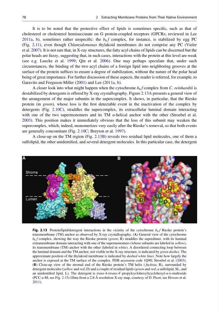
destabilized by detergents is offered by X-ray crystallography. Figure 2.13A presents a general view of
the arrangement of the major subunits in the supercomplex. It shows, in particular, that the Rieske
protein (in green), whose loss is the first detectable event in the inactivation of the complex by
detergents (Fig. 2.10C), straddles the supercomplex, its extracellular luminal domain interacting
with one of the two supermonomers and its TM α-helical anchor with the other (Stroebel et al.
2003). This position makes it immediately obvious that the loss of this subunit may weaken the
supercomplex, which, indeed, monomerizes very easily after the Rieske’s removal, so that both events
are generally concomitant (Fig. 2.10C; Breyton et al. 1997).
A close-up on the TM region (Fig. 2.13B) reveals two residual lipid molecules, one of them a
sulfolipid, the other unidentified, and several detergent molecules. In this particular case, the detergent
Fig. 2.13 Protein/lipid/detergent interactions in the vicinity of the cytochrome b 6 f Rieske protein’s
transmembrane (TM) anchor as observed by X-ray crystallography. (A) General view of the cytochrome
b 6 f complex, showing the way the Rieske protein (green; R) straddles the superdimer, with its luminal
extramembrane domain interacting with one of the supermonomers (whose subunits are labeled in yellow),
its transmembrane (TM) anchor with the other (labeled in white). A disordered connecting loop between
the luminal domain and the TM anchor, not visible in the X-ray structure, is indicated by green dashes. The
approximate position of the thylakoid membrane is indicated by dashed white lines. Note how largely the
anchor is exposed at the TM surface of the complex. PDB accession code 1Q90; Stroebel et al. (2003).
(B) Close-up view of the stromal end of the Rieske protein’s TM helix ( fuchsia; R), surrounded by
detergent molecules (yellow and red; D) and a couple of residual lipids (green and red; a sulfolipid, SL, and
an unidentified lipid, L). The detergent is trans-4-(trans-4
0 -propylcyclohexyl)cyclohexyl-α-D-maltoside
(PCC-a-M; see Fig. 2.15) (Data from a 2.8-Å resolution X-ray map, courtesy of D. Picot; see Hovers et al.
2011).
78
2 Extracting Membrane Proteins from Their Native Environment
cholesterol or cholesterol hemisuccinate on G protein-coupled receptors (GPCRs; reviewed in Lee
2011a, b), sometimes rather unspecific: the b 6 f complex, for instance, is stabilized by egg PC
(Fig. 2.11), even though Chlamydomonas thylakoid membranes do not comprise any PC (Vieler
et al. 2007). It is not rare that, in X-ray structures, the fatty acyl chains of lipids can be discerned but the
polar heads are fuzzy, suggesting that, in such cases, interactions with the protein at this level are weak
(see e.g. Luecke et al. 1999; Qin et al. 2006). One may perhaps speculate that, under such
circumstances, the binding of the two acyl chains of a foreign lipid into neighboring grooves at the
surface of the protein suffices to ensure a degree of stabilization, without the nature of the polar head
being of great importance. For further discussion of these aspects, the reader is referred, for example, to
Garavito and Ferguson-Miller (2001) and Lee (2011a, b).
A closer look into what might happen when the cytochrome b 6 f complex from C. reinhardtii is
destabilized by detergents is offered by X-ray crystallography. Figure 2.13A presents a general view of
the arrangement of the major subunits in the supercomplex. It shows, in particular, that the Rieske
protein (in green), whose loss is the first detectable event in the inactivation of the complex by
detergents (Fig. 2.10C), straddles the supercomplex, its extracellular luminal domain interacting
with one of the two supermonomers and its TM α-helical anchor with the other (Stroebel et al.
2003). This position makes it immediately obvious that the loss of this subunit may weaken the
supercomplex, which, indeed, monomerizes very easily after the Rieske’s removal, so that both events
are generally concomitant (Fig. 2.10C; Breyton et al. 1997).
A close-up on the TM region (Fig. 2.13B) reveals two residual lipid molecules, one of them a
sulfolipid, the other unidentified, and several detergent molecules. In this particular case, the detergent
Fig. 2.13 Protein/lipid/detergent interactions in the vicinity of the cytochrome b 6 f Rieske protein’s
transmembrane (TM) anchor as observed by X-ray crystallography. (A) General view of the cytochrome
b 6 f complex, showing the way the Rieske protein (green; R) straddles the superdimer, with its luminal
extramembrane domain interacting with one of the supermonomers (whose subunits are labeled in yellow),
its transmembrane (TM) anchor with the other (labeled in white). A disordered connecting loop between
the luminal domain and the TM anchor, not visible in the X-ray structure, is indicated by green dashes. The
approximate position of the thylakoid membrane is indicated by dashed white lines. Note how largely the
anchor is exposed at the TM surface of the complex. PDB accession code 1Q90; Stroebel et al. (2003).
(B) Close-up view of the stromal end of the Rieske protein’s TM helix ( fuchsia; R), surrounded by
detergent molecules (yellow and red; D) and a couple of residual lipids (green and red; a sulfolipid, SL, and
an unidentified lipid, L). The detergent is trans-4-(trans-4
0 -propylcyclohexyl)cyclohexyl-α-D-maltoside
(PCC-a-M; see Fig. 2.15) (Data from a 2.8-Å resolution X-ray map, courtesy of D. Picot; see Hovers et al.
2011).
78
2 Extracting Membrane Proteins from Their Native Environment