

micelles. The CMC of pure HG is variously quoted to lie between ~16.5 mM (Ruiz et al.
1994), ~19.5 mM (Enzo Life Science web site), and 20–25 mM (Vegatec web site). Given
that the concentration of free monomers drops somewhat in the presence of lipids, one can
estimate that the concentration of micellar detergent was in the range of 30–35 mM in the
presence of 50 mM HG (left) and of 80–85 mM in the presence of 100 mM HG (right). The
rate of inactivation was faster in the second case, with, for instance, after 30 min and at a 1:10
PC/HG ratio, ~1/3 inactivation at 50 mM HG vs. ~1/2 at 100 mM. Under these two sets of
conditions, the composition of the micelles is virtually identical, so the environment the
complex experiences has the same physical properties. A change in those cannot be invoked
to explain the faster inactivation at 100 mM HG.
• Under these three sets of circumstances, what does change with the concentration of
surfactants is the ratio of detergent to protein and, if applicable, to lipids: as this ratio
increases, any equilibrium between protein/protein, protein/lipid, protein/detergent, lipid/
lipid, and lipid/detergent interactions becomes displaced in favor of the detergent. This can
affect, directly or indirectly, interactions between TM protein segments, whether within
subunits or between subunits. Lipid molecules and protein subunits that interact to stabilize
the complex will tend to be diluted in the micelles and replaced by detergent. Detergent
molecules are, as a rule, smaller than lipid ones, and, with some exceptions like the LMNG
series (see Fig. 2.15), they usually comprise a single hydrophobic chain. As a consequence,
(i) they may not have the same “clamping” effect that a two-chain lipid may have when it
straddles two TM segments or subunits, and (ii) they may more easily intrude into cranks in
the protein structure, weakening protein/protein interactions and extracting bound lipids and
cofactors (cf. Chap. 1, § 1.5.2). The protective effect of lipids, in turn, may be due (i) to
stabilizing interactions they establish with the protein and (ii) to their blocking or slowing
down the intrusion of detergent molecules into protein cranks or clefts.
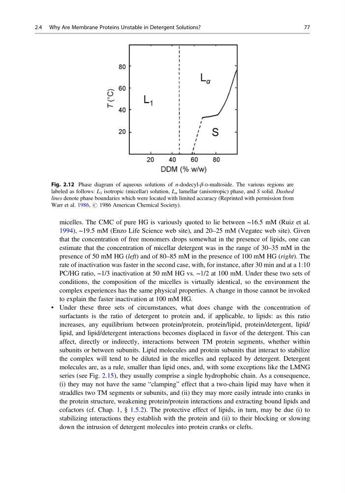
Fig. 2.12 Phase diagram of aqueous solutions of n-dodecyl-β-D-maltoside. The various regions are
labeled as follows: L 1 isotropic (micellar) solution, L α lamellar (anisotropic) phase, and S solid. Dashed
lines denote phase boundaries which were located with limited accuracy (Reprinted with permission from
Warr et al. 1986, # 1986 American Chemical Society).
2.4 Why Are Membrane Proteins Unstable in Detergent Solutions?
77
1994), ~19.5 mM (Enzo Life Science web site), and 20–25 mM (Vegatec web site). Given
that the concentration of free monomers drops somewhat in the presence of lipids, one can
estimate that the concentration of micellar detergent was in the range of 30–35 mM in the
presence of 50 mM HG (left) and of 80–85 mM in the presence of 100 mM HG (right). The
rate of inactivation was faster in the second case, with, for instance, after 30 min and at a 1:10
PC/HG ratio, ~1/3 inactivation at 50 mM HG vs. ~1/2 at 100 mM. Under these two sets of
conditions, the composition of the micelles is virtually identical, so the environment the
complex experiences has the same physical properties. A change in those cannot be invoked
to explain the faster inactivation at 100 mM HG.
• Under these three sets of circumstances, what does change with the concentration of
surfactants is the ratio of detergent to protein and, if applicable, to lipids: as this ratio
increases, any equilibrium between protein/protein, protein/lipid, protein/detergent, lipid/
lipid, and lipid/detergent interactions becomes displaced in favor of the detergent. This can
affect, directly or indirectly, interactions between TM protein segments, whether within
subunits or between subunits. Lipid molecules and protein subunits that interact to stabilize
the complex will tend to be diluted in the micelles and replaced by detergent. Detergent
molecules are, as a rule, smaller than lipid ones, and, with some exceptions like the LMNG
series (see Fig. 2.15), they usually comprise a single hydrophobic chain. As a consequence,
(i) they may not have the same “clamping” effect that a two-chain lipid may have when it
straddles two TM segments or subunits, and (ii) they may more easily intrude into cranks in
the protein structure, weakening protein/protein interactions and extracting bound lipids and
cofactors (cf. Chap. 1, § 1.5.2). The protective effect of lipids, in turn, may be due (i) to
stabilizing interactions they establish with the protein and (ii) to their blocking or slowing
down the intrusion of detergent molecules into protein cranks or clefts.
Fig. 2.12 Phase diagram of aqueous solutions of n-dodecyl-β-D-maltoside. The various regions are
labeled as follows: L 1 isotropic (micellar) solution, L α lamellar (anisotropic) phase, and S solid. Dashed
lines denote phase boundaries which were located with limited accuracy (Reprinted with permission from
Warr et al. 1986, # 1986 American Chemical Society).
2.4 Why Are Membrane Proteins Unstable in Detergent Solutions?
77