

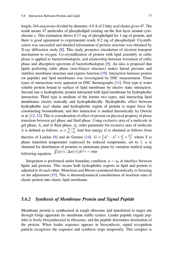
length, 244 angstroms divided by diameter, 4.8 Å of 2 fatty acyl chains gives 47. The
result means 47 molecules of phospholipid existing on the first layer around cytochrome c. This estimation shows 0.17 mg of phospholipid for 1 mg of protein, and
there is good agreement to experimental result, 0.2 mg of phospholipid. Crystallization was succeeded and detailed information of protein structure was obtained by
X-ray diffraction study [8]. This study promotes elucidation of electron transport
mechanism to oxygen. Co-crystallization of protein with lipid assembly in cubic
phase is applied to bacteriorhodopsin, and relationship between formation of cubic
phase and absorption spectrum of bacteriorhodopsin [9]. An idea is proposed that
lipids preferring cubic phase (non-bilayer structure) makes lateral pressure and
stabilize membrane structure and express function [10]. Interaction between protein
(or peptide) and lipid membranes was investigated by DSC measurement. Three
types of interactions were appeared on DSC thermographs [11]. First type is water
soluble protein bound to surface of lipid membrane by electro static interaction.
Second one is hydrophobic protein interacted with lipid membrane by hydrophobic
interaction. Third type is medium of the former two types, and interacting lipid
membranes electro statically and hydrophobically. Hydrophobic effect between
hydrophobic acyl chains and hydrophobic region of protein is major force for
constructing biomembrane, and this interaction is studied theoretically by Owicki
et al. [12, 13]. This is consideration of effect of protein on physical property of phase
transition between gel phase and fluid phase. Using exclusive area of a molecule in
gel phase, A s and in fluid phase, A f, order parameter for excusive area of molecule
A is defined as follows. u
A f ÀA
A f ÀA s
And free energy G is obtained as follows from
theories of Landau [9] and de Gennes [14]. G ¼
T
2 u
2
À u
3
þ
u
4
2 þ
u
j j
2
2 where T is
phase transition temperature expressed by reduced temperature, set to 1. u is
obtained for distribution of proteins in membrane plane by variation method using
following equation.
RR
u r
ð Þ; Δu r
ð Þ
j
j
j
j dr
2 r ¼ min .
Integration is performed under boundary condition, u ¼ u 0 at interface between
lipids and proteins. This means both hydrophobic regions in lipid and protein is
adjusted to fit each other. Mouritsen and Bloom considered theoretically in focusing
on the adjustment [15]. This is thermodynamical consideration of insertion state of
elastic protein into elastic lipid membrane.
5.6.2 Synthesis of Membrane Protein and Signal Peptide
Membrane protein is synthesized in rough ribosome and transferred to target site
through Golgi apparatus by membrane traffic system. Leader peptide (signal peptide) is firstly biosynthesized in ribosome, and the peptide determines destination of
the protein. When leader sequence appears in biosynthesis, signal recognition
particle recognizes the sequence and synthesis stops temporally. This complex is
5.6 Interaction Between Protein and Lipid
77
result means 47 molecules of phospholipid existing on the first layer around cytochrome c. This estimation shows 0.17 mg of phospholipid for 1 mg of protein, and
there is good agreement to experimental result, 0.2 mg of phospholipid. Crystallization was succeeded and detailed information of protein structure was obtained by
X-ray diffraction study [8]. This study promotes elucidation of electron transport
mechanism to oxygen. Co-crystallization of protein with lipid assembly in cubic
phase is applied to bacteriorhodopsin, and relationship between formation of cubic
phase and absorption spectrum of bacteriorhodopsin [9]. An idea is proposed that
lipids preferring cubic phase (non-bilayer structure) makes lateral pressure and
stabilize membrane structure and express function [10]. Interaction between protein
(or peptide) and lipid membranes was investigated by DSC measurement. Three
types of interactions were appeared on DSC thermographs [11]. First type is water
soluble protein bound to surface of lipid membrane by electro static interaction.
Second one is hydrophobic protein interacted with lipid membrane by hydrophobic
interaction. Third type is medium of the former two types, and interacting lipid
membranes electro statically and hydrophobically. Hydrophobic effect between
hydrophobic acyl chains and hydrophobic region of protein is major force for
constructing biomembrane, and this interaction is studied theoretically by Owicki
et al. [12, 13]. This is consideration of effect of protein on physical property of phase
transition between gel phase and fluid phase. Using exclusive area of a molecule in
gel phase, A s and in fluid phase, A f, order parameter for excusive area of molecule
A is defined as follows. u
A f ÀA
A f ÀA s
And free energy G is obtained as follows from
theories of Landau [9] and de Gennes [14]. G ¼
T
2 u
2
À u
3
þ
u
4
2 þ
u
j j
2
2 where T is
phase transition temperature expressed by reduced temperature, set to 1. u is
obtained for distribution of proteins in membrane plane by variation method using
following equation.
RR
u r
ð Þ; Δu r
ð Þ
j
j
j
j dr
2 r ¼ min .
Integration is performed under boundary condition, u ¼ u 0 at interface between
lipids and proteins. This means both hydrophobic regions in lipid and protein is
adjusted to fit each other. Mouritsen and Bloom considered theoretically in focusing
on the adjustment [15]. This is thermodynamical consideration of insertion state of
elastic protein into elastic lipid membrane.
5.6.2 Synthesis of Membrane Protein and Signal Peptide
Membrane protein is synthesized in rough ribosome and transferred to target site
through Golgi apparatus by membrane traffic system. Leader peptide (signal peptide) is firstly biosynthesized in ribosome, and the peptide determines destination of
the protein. When leader sequence appears in biosynthesis, signal recognition
particle recognizes the sequence and synthesis stops temporally. This complex is
5.6 Interaction Between Protein and Lipid
77