

different membrane fluidity. Hinge part of high fluidity indicates DMPC rich region
and straight part of low fluidity indicates DMPE rich region (Fig. 3.21).
3.7.5 Optical Microscopy Using Evanescent-Field
In the preceding sections, methodologies that enable visualization of the physical
properties, eg., fluidity of biomembranes are presented. In this section, a method for
visualization of the phenomena occurring at the glass-water interface, where cultured
cells are bound or molecules in the solution interact with molecules bound to the
interface. Figure 3.22a shows the generation of an evanescent wave, a form of nearfield illumination. A laser beam which is incident upon the interface of glass and
water with the incident angle equal to or larger than the angle called critical angle
(~61 degrees), is totally internally reflected, which is accompanied by a generation at
II-CCD
DM
DM
DM
M
M
original image plane
SL
MF(440nm)
MF(490nm)
Lamp
ND
L
OL
L
L
temperature-controlled stage
epifluorescence microscope
(AxioPlan)
Sample
Dual-view optics
ExF
EmF
QWP
Peltier
Control
motor-drive Z controller
Video
Recorder
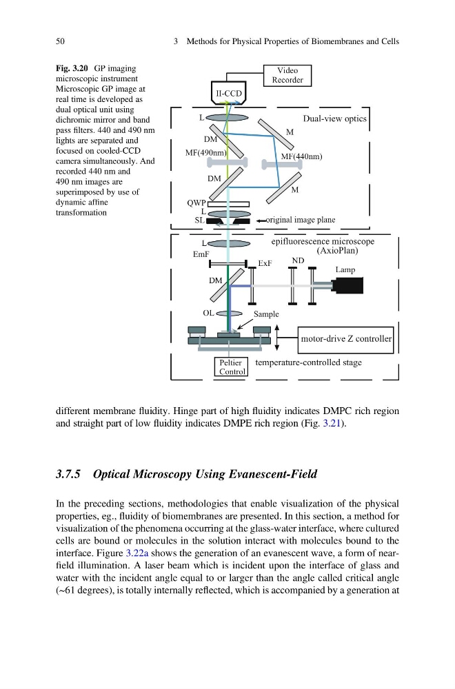
Fig. 3.20 GP imaging
microscopic instrument
Microscopic GP image at
real time is developed as
dual optical unit using
dichromic mirror and band
pass filters. 440 and 490 nm
lights are separated and
focused on cooled-CCD
camera simultaneously. And
recorded 440 nm and
490 nm images are
superimposed by use of
dynamic affine
transformation
50
3 Methods for Physical Properties of Biomembranes and Cells
and straight part of low fluidity indicates DMPE rich region (Fig. 3.21).
3.7.5 Optical Microscopy Using Evanescent-Field
In the preceding sections, methodologies that enable visualization of the physical
properties, eg., fluidity of biomembranes are presented. In this section, a method for
visualization of the phenomena occurring at the glass-water interface, where cultured
cells are bound or molecules in the solution interact with molecules bound to the
interface. Figure 3.22a shows the generation of an evanescent wave, a form of nearfield illumination. A laser beam which is incident upon the interface of glass and
water with the incident angle equal to or larger than the angle called critical angle
(~61 degrees), is totally internally reflected, which is accompanied by a generation at
II-CCD
DM
DM
DM
M
M
original image plane
SL
MF(440nm)
MF(490nm)
Lamp
ND
L
OL
L
L
temperature-controlled stage
epifluorescence microscope
(AxioPlan)
Sample
Dual-view optics
ExF
EmF
QWP
Peltier
Control
motor-drive Z controller
Video
Recorder
Fig. 3.20 GP imaging
microscopic instrument
Microscopic GP image at
real time is developed as
dual optical unit using
dichromic mirror and band
pass filters. 440 and 490 nm
lights are separated and
focused on cooled-CCD
camera simultaneously. And
recorded 440 nm and
490 nm images are
superimposed by use of
dynamic affine
transformation
50
3 Methods for Physical Properties of Biomembranes and Cells